Back to Journals » Drug Design, Development and Therapy » Volume 20
Formulation Progress, Challenges, and Perspectives of Anti-Inflammatory Natural Products
Authors Fang H ![]() , Wu A, Zhao M
, Wu A, Zhao M ![]() , Nan X, Yang L, Liu H
, Nan X, Yang L, Liu H ![]()
Received 3 March 2026
Accepted for publication 29 April 2026
Published 7 May 2026 Volume 2026:20 606531
DOI https://doi.org/10.2147/DDDT.S606531
Checked for plagiarism Yes
Review by Single anonymous peer review
Peer reviewer comments 3
Editor who approved publication: Professor Anastasios Lymperopoulos
Hongran Fang,1,* Ailing Wu,2,* Muxin Zhao,1,* Xichen Nan,1 Luhan Yang,1 Hao Liu1
1School of Pharmacy, Southwest Medical University, Luzhou, Sichuan, People’s Republic of China; 2Department of Anesthesiology, The Second People’s Hospital of Neijiang, Southwest Medical University, Neijiang, Sichuan, People’s Republic of China
*These authors contributed equally to this work
Correspondence: Hao Liu, School of Pharmacy, Southwest Medical University, No. 1 Section 1, Xiang Lin Road, Longmatan District, Luzhou, Sichuan, 646000, People’s Republic of China, Email [email protected]
Abstract: Inflammation is a complex and highly regulated defensive response of the body to injury, infection, or abnormal stimuli (such as pathogens, toxins, and physical/chemical damage). Natural anti-inflammatory drugs hold significant potential in the pharmaceutical field due to their multi-target effects, high safety profiles, and low toxicity. For example, EGCG can inhibit the phosphorylation of p38 and JNK, thereby reducing the activation of the AP-1 transcription factor and subsequently downregulating the expression of inflammatory genes. Luteolin inhibits inflammasome assembly by blocking potassium ion efflux, suppressing mitochondrial reactive oxygen species generation, or directly binding to NLRP3. Over the past decade, extensive research has been conducted on their physicochemical properties and dosage forms, leading to the development of various natural anti-inflammatory drug formulations using both traditional and modern technologies. Furthermore, the combination of these natural anti-inflammatory agents with other drugs can further expand their therapeutic applications. Meanwhile, emerging technologies such as 3D printing and AI-assisted design have demonstrated significant potential for application in formulation development. Despite these advancements, the current research field still faces critical challenges: the use of toxic excipients in certain formulations poses biosafety risks (for example, glutaraldehyde offers advantages such as high cross-linking efficiency, strong cross-linking strength, and mature manufacturing processes, but it also exhibits high toxicity and biosafety deficiencies); and the translation of research findings on natural anti-inflammatory drugs into commercial products remains insufficient (due to challenges in large-scale production, storage difficulties, and regulatory standards). Through this review, we hope to draw more attention to the development potential of natural anti-inflammatory drugs and the aforementioned issues, as well as to offer some foresight for the exploration and development of other natural products. An infographic showing a workflow from preparation technologies to dosage forms and clinical application sites. Preparation methods are labeled Microfluidic technology and Electro-spray technology, illustrated by a microfluidic chip and a lab apparatus with a droplet. Preparation leads to two dosage forms labeled Tablets and Microparticles, illustrated by three round tablets and three textured spheres. Application is shown with a human body silhouette and organ callouts listing target conditions: eye, Conjunctivitis, Keratitis, Blepharitis ciliaris; nasal and sinus, Sinusitis, Nasal vestibulitis, Rhinitis; oral and dental, Periodontitis, Pulpitis, Gingivitis; throat and stomach, Gastritis, Ulcerative, Tonsillitis; intestine, Ulcerative colitis, Norovirus enteritis; anorectal, Hemorrhoid, Anal fistula, Perianal abscess; skin, Eczema, Psoriasis, Pityriasis rosea.An infographic on microfluidic and electro-spray preparation of tablets and microparticles for diseases treatment.
Keywords: inflammation, natural product, dosage forms, drug delivery, 3D printing, artificial intelligence
Introduction
Inflammation is a protective immune response of an organism to injury, infection, or other stimuli,1,2 that can be triggered by a variety of external and internal factors, leading to either acute or chronic disease. Acute inflammation progresses rapidly, with a short golden treatment window, which can easily lead to organ-specific damage and significantly increase the risk of lifelong chronic diseases and cancer. Acute inflammation-related diseases account for nearly 20% of all deaths worldwide, with mortality rates reaching up to 50% in resource-limited areas, constituting a major global public health crisis.3 Meanwhile, chronic inflammatory diseases collectively constitute a major global cause of death, accounting for 20% of all cancer-related deaths. Additionally, chronic inflammation has been implicated in developing various critical diseases, such as diabetes, Alzheimer’s disease, and even some types of cancer, are included.4,5 The detrimental effects of inflammation should not be underestimated.
Anti-inflammatory natural products are bioactive compounds extracted from natural organisms, capable of inhibiting inflammatory mediators, inflammatory pathways, or inflammatory responses, thereby alleviating acute or chronic inflammation without the need for synthetic chemical modifications. Multiple drugs can achieve the goal of regulating the inflammatory response and maintaining the balance between pro-inflammatory and anti-inflammatory processes, including nonsteroidal anti-inflammatory drugs (NSAIDs), glucocorticoid analogs, and natural anti-inflammatory agents. However, long-term use of NSAIDs and glucocorticoid analogs may lead to a series of adverse reactions and drug resistance in humans. For example, the adverse effects of aspirin primarily involve gastrointestinal damage,6 bleeding risk,7 and hepatorenal injury.8 Prednisone may lead to insulin resistance and hyperglycemia,9 osteoporosis and fractures.10 Compared to synthetic NSAIDs and glucocorticoids, anti-inflammatory natural products exhibit superior advantages in long-term medication safety and resistance management. For instance, existing studies have conducted comparative analyses, demonstrating that curcumin (Cur) and boswellia extract exhibit efficacy comparable to NSAIDs in the treatment of osteoarthritis with lower adverse effects. Long-term use is recommended.11–14 Some studies have compared andrographolide and NSAIDs in terms of anti-inflammatory efficacy and immunosuppressive effects,15 as well as Vitamin C and glucocorticoids in terms of therapeutic outcomes and withdrawal rebound.16 The results consistently indicated that natural anti-inflammatory compounds serve as effective alternatives to the aforementioned synthetic drugs, making them better for treating long-term inflammatory diseases.17,18
Up to now, studies on the chemical structure,19,20 and anti-inflammatory activity21–23 of anti-inflammatory natural products have been reviewed in detail elsewhere. Guo et al systematically summarized the structures of over 260 anti-inflammatory natural products from the past two decades and discussed their structure-activity relationships in anti-inflammatory effects.19 Choo et al classified anti-inflammatory natural products according to their chemical skeletons and discussed the essential groups required for exerting anti-inflammatory activity.20 Allijn et al summarized the anti-inflammatory activities of 102 plant-derived natural products across 25 common assays and evaluated their inhibitory effects on inflammatory mediators, oxidative stress.21 Peng et al and Zheng et al respectively discussed the anti-inflammatory activities of saponin-based anti-inflammatory natural products and various common anti-inflammatory natural products.22,23
Despite the promising anti-inflammatory activity and biological safety of natural products, their application are severely limited by inherent physicochemical drawbacks. Most anti-inflammatory natural products exhibit poor water solubility, insufficient chemical stability, low oral bioavailability, rapid metabolic elimination, and lack of targeted delivery to inflammatory sites. These disadvantages directly lead to weak in vivo efficacy, high effective doses, and unsatisfactory therapeutic outcomes. In addition, the successfully prepared anti-inflammatory natural product formulations still face the issue of insufficient translation of research findings into commercial products. The reasons for insufficient translation include not only defects in the physicochemical properties of the drug itself, but also challenges in large-scale production, formulation storage difficulties, insufficient clinical evidence, and regulatory standard issues. Lab-scale preparation techniques are difficult to scale up due to high costs, strict process control requirements, and poor batch-to-batch consistency. The instability of nanoformulations during storage and transportation further shortens the shelf life and increases supply chain burdens. In addition, unstable raw material sources, complex regulatory approval pathways, huge clinical evaluation costs, and insufficient clinical evidence collectively hinder industrialization. These multi-dimensional bottlenecks make it difficult to transform advanced formulation strategies into safe, effective, and economically feasible commercial anti-natural products.
Moreover, some marketed or patented anti-inflammatory natural preparations exhibit the drawbacks of solvent toxicity and toxic excipients, which may raise safety problems. For instance, glutaraldehyde is commonly used as a cross-linking agent in drug preparation. Although glutaraldehyde exhibits high cross-linking efficiency and significant cross-linking effects, its potential toxicity and carcinogenicity have posed biosafety concerns. Therefore, the development of rational drug delivery systems and optimized formulations has become an essential strategy to address these bottlenecks.
In view of the above dilemmas, this review focuses on the formulation development of anti-inflammatory natural products. We systematically summarize the progress in delivery systems, including nanoparticles, liposomes, micelles, microcapsules, and other advanced formulations for enhancing solubility, stability, bioavailability, and targeted anti-inflammatory effects. We also analyzed the integration of natural pharmaceutical formulations with novel technologies, such as 3D printing and computer-aided design (CAD), and the obstacles and pharmaceutical development approaches for the use of natural anti-inflammatory drugs in combination with other drugs. We also analyze the critical challenges in scale-up production, storage stability, quality control, biosafety, and clinical translation. Finally, we prospect the future directions of intelligent delivery, targeted therapy, green preparation, and industrial translation. This review aims to offer valuable references for accelerating the rational design, clinical application, and commercialization of anti-inflammatory natural product formulations.
Mechanism of Inflammation and Pharmacological Actions of Natural Anti-Inflammatory Products
Inflammation and Oxidative Stress
Inflammation is a complex pathophysiological process initiated when the body is exposed to endogenous or exogenous damaging factors (such as pathogens, trauma, toxins, or ischemia). In this process, pattern recognition receptors (PRRs) detect danger signals (PAMPs/DAMPs), triggering a coordinated response involving the vascular, immune, and nervous systems.24
The triggering mechanism of acute inflammatory responses induced by infection or tissue injury relies on PRRs recognizing pathogen-associated molecular patterns (PAMPs) and damage-associated molecular patterns (DAMPs), particularly Toll-like receptors (TLRs) and nucleotide-binding oligomeric domain-like receptors (NLRs).
The initial recognition process of acute inflammation is mediated by tissue-resident macrophages and mast cells, which subsequently trigger the production of various inflammatory mediators, including chemokines, cytokines, vasoactive amines, eicosanoids, and products of proteolytic cascades. These mediators promote the activation of vascular endothelial cells, transiently increase vascular permeability, and selectively mediate the recruitment of inflammatory cells such as neutrophils and monocytes to the injury site, while simultaneously inhibiting the excretion of erythrocytes. This selectivity is achieved by inducing ligation of endothelial-cell selectins with integrins and chemokine receptors on leukocytes. Neutrophils kill invading pathogens by releasing reactive oxygen species (ROS) and reactive nitrogen species, protease 3, and cathepsin G.25,26 Mast cells can release mediators to regulate immune responses and promote inflammatory diseases while avoiding catastrophic anaphylactic shock. They can secrete specific cytokines (TNF-α, IL-4, IL-5, IL-13) or growth factors (vascular endothelial growth factor, fibroblast growth factor), influencing local immunity by promoting tissue repair during inflammation or modulating the balance between pro-inflammatory and anti-inflammatory signaling pathways.27
If the acute inflammatory response fails to eliminate the pathogen, the neutrophil infiltration is replaced by macrophages, and in cases of infection, by T cells. If the combined effects of these cells remain insufficient, a chronic inflammatory state will occur. The typical features of chronic inflammation include persistent infiltration of inflammatory cells, prolonged excessive release of pro-inflammatory mediators, continuously enhanced oxidative stress response, and abnormal activation of tissue repair-related signaling pathways. A persistent inflammatory state induces cytokine storms, exacerbates oxidative damage, leads to excessive degradation of the extracellular matrix, disrupts collagen deposition, and causes abnormal apoptosis, resulting in sustained damage to local tissues, cells, and organ structures.28,29
Oxidative stress and inflammation mutually drive each other.30 The OS results from the overproduction of ROS in living cells. Oxidative stress induces inflammation by activating inflammation-related signaling pathways, such as NF-κB and MAPK, and by promoting the release of pro-inflammatory factors (eg, TNF-α, IL-6, IL-1β). Inflammation, in turn, exacerbates oxidative stress. Inflammatory cells, including neutrophils and macrophages, release substantial amounts of ROS (eg, superoxide anion, hydrogen peroxide) during pathogen clearance, further exacerbating oxidative stress.31,32 Thus, the synergistic anti-inflammatory and antioxidant effects are crucial for mitigating inflammatory responses in critical illnesses. The synergistic anti-inflammatory and antioxidant effects.
Action Mechanism of Natural Anti-Inflammatory Products
The advantages of natural anti-inflammatory compounds extend beyond the previously mentioned higher long-term safety profile and reduced susceptibility to hormone dependence and typical drug resistance. Additionally, they exhibit multi-target, multi-pathway characteristics, which distinctly differentiate them from single-target synthetic drugs in anti-inflammatory therapy. The multi-functional pathways of natural anti-inflammatory products are intricately interconnected, collectively regulating the intensity and duration of inflammatory responses.
The NF-κB pathway is the most critical regulatory target for natural anti-inflammatory products. This pathway is activated by inflammatory stimuli, leading to receptor activation and recruitment of adapter proteins (such as TRADD and RIP), which further activate IκB kinase (IKK). IKK phosphorylates IκB, resulting in its degradation, enabling nuclear translocation of NF-κB (p50/p65) and then initiating transcription of pro-inflammatory genes.33 For example, the polyphenolic component quercetin inhibits IκB degradation, thereby blocking p65 nuclear translocation and suppressing inflammatory responses.34
The MAPK pathway comprises three parallel cascaded pathways: p38 kinase, c-Jun N-terminal kinase (JNK), and extracellular regulatory kinase (ERK). ERK is primarily involved in cell proliferation and occasionally participates in inflammation, whereas JNK and p38 are mainly associated with inflammation and apoptosis.35 The ERK pathway induces the expression of anti-inflammatory factors such as IL-10; the JNK pathway dominates AP-1 transcriptional activation, driving the expression of TNF-α, IL-2, and MMPs; while the p38 pathway induces the expression of TNF-α, IL-1β, IL-6, COX-2, and iNOS.36,37 For example, the natural product epigallocatechin gallate (EGCG) reduces the activation of the AP-1 transcription factor by inhibiting p38 and JNK phosphorylation, thereby downregulating the expression of inflammatory genes.38
MAPK and NF-κB do not operate independently; there is synergy and overlap between them, which can form a positive feedback loop. TAK1 can simultaneously activate the IKK complex (NF-κB) and the MKK3/6-JNK/p38 (MAPK) pathway, enabling parallel activation of both pathways.39 Phosphorylated p38 can activate MSK1/2 and phosphorylate transcription factors (CREB, ATF-1), which synergistically enhance the expression of inflammatory genes in collaboration with NF-κB.40 Natural products such as EGCG can simultaneously inhibit NF-κB nuclear translocation and p38 phosphorylation, achieving dual pathway blockade.41
The JAK-STAT pathway serves as a critical integration node for both pro-inflammatory and anti-inflammatory signaling pathways. It originates from two key molecular classes: Janus kinases (JAKs) and signal transducer and transcription activator factors (STATs). Cytokines (such as interferon (IFN) and interleukins (IL-2/6/12)) induce receptors dimerization, subsequently triggering mutual phosphorylation and activation of the JAK kinases (JAK1/2/3 and Tyk2) bound to the intracellular domain. The activated JAK subsequently phosphorylates specific tyrosine residues in the intracellular domain of the receptors, providing docking sites for STAT proteins. After binding to the phosphorylated receptor, STAT is phosphorylated by JAK. Phosphorylated STAT forms homologous or heterologous dimers, subsequently translocates into the nucleus, and binds to specific DNA sequences, thereby initiating the transcription of target genes.42,43 Natural anti-inflammatory components such as curcumin inhibit JAK1/2 phosphorylation and block STAT3 nuclear translocation.44 Berberine inhibits JAK kinase activity, downregulates the expression of JAK2 and STAT3, blocks the phosphorylation and dimerization of STAT proteins, and interferes with signal transduction pathways involving IFN-γ and IL-6.45
JAK-STAT exhibits both overlap and synergy with NF-κB and MAPK. TNF-α activates NF-κB, which induces IL-6 expression. IL-6 then promotes the expression of survival genes (Bcl-xL, Mcl-1) via JAK-STAT3, thereby conferring apoptosis resistance to inflammatory cells. JAK activation can indirectly activate ERK; conversely, ERK can phosphorylate STAT3, enhancing its transcriptional activity.
NLRP3 inflammasomes regulate the maturation and secretion of pro-inflammatory cytokines, participating in the pathogenesis of various chronic inflammatory diseases.46 When activated by various stimuli, such as ROS, NLRP3 binds to the adapter protein ASC and pro-caspase-1, leading to the autocatalytic activation of caspase-1. This process subsequently cleaves pro-IL-1β and pro-IL-18 into their bioactive forms, IL-1β and IL-18, thereby enhancing the inflammatory response.47 Natural products such as luteolin inhibit inflammatory small body assembly by blocking potassium efflux, suppressing mitochondrial reactive oxygen species generation, or directly binding to NLRP3.48
Activation of the Nrf2/ARE antioxidant pathway represents another critical mechanism. Under oxidative stress conditions, this process promotes the dissociation and nuclear translocation of Nrf2 from Keap1, enabling its binding to the antioxidant response element (ARE). This induces the expression of protective proteins such as HO-1, NQO1, and GST, thereby scavenging reactive oxygen species and suppressing oxidation-stress-driven inflammation.
The PPAR-γ nuclear receptor pathway reduces the expression of M1 polarization markers (iNOS) by inhibiting STAT-1 phosphorylation, while promoting STAT-6 phosphorylation to increase the expression of M2 markers (Arg-1, Fizz 1, Ym 1).49 PPAR-γ occupies a central node in metabolic-inflammation regulation, forming complex interactions with multiple pathways: it exhibits antagonistic equilibrium with NF-κB, synergistic effects with Nrf2/ARE, cross-regulation with AMPK, and negative modulation with NLRP3 inflammasome. The PI3K/Akt/mTOR pathway regulates cell survival, metabolism, and autophagy. PI3K/Akt/mTOR does not operate in isolation but forms a complex network with multiple inflammatory pathways. The cross-regulation of these pathways endows natural anti-inflammatory products with multi-target synergistic effects, enabling rapid suppression of acute inflammation while also offering unique advantages in chronic inflammation management by promoting the synthesis of pro-inflammatory resolution mediators.
Pharmaceutical Designs for the Application of a Single Natural Anti-Inflammatory Drug
Nano-Preparations
Lipid-Based Nanocarriers
Current pharmacotherapy primarily centers on monotherapy, employing various drug delivery systems to achieve optimal therapeutic efficacy. Lipid-based nanocarriers mainly encompass liposomes, solid lipid nanoparticles (SLNs), and nanostructured lipid carriers (NLCs). Their fundamental advantages reside in the exceptional biocompatibility and delivery efficacy imparted by the combination of lipids and nanostructures. Depending on specific therapeutic requirements and drug properties, drug-loaded liposomes can be modified using various polymer materials to form composite liposomes (comprising polymers, targeting molecules, environment-responsive components, and pH/temperature-sensitive lipids, etc). This approach not only allows liposomes to carry drugs but also possesses additional functionalities, such as prolonged circulation, targeted delivery, and stimulus-responsive responses, as illustrated in Figure 1.
 |
Figure 1 Schematic diagram of the preparation of polymeric composite liposomes (PMCL) loaded with anti-inflammatory drugs and animal cell experiments. Anti-inflammatory drugs and phospholipids are combined to form drug-loaded liposomes, which are further modified with polymers (polysaccharides, peptides, etc.) to create Polymer-modified Complex Liposomes. The anti-inflammatory efficacy of these liposomes can be validated in animal models. |
The inimitable structure of liposomes enables encapsulation of both hydrophilic and hydrophobic drug components. However, their high sensitivity to environmental factors such as pH, ionic strength, and temperature limits their application in many anti-inflammatory drug delivery systems. Given that glycolipids, glycoproteins, and proteins in cell membranes enhance stability and functionality in fluid-mosaic membrane models, liposomes can be modified by attaching polysaccharides or proteins to their surfaces to alter their properties. Cheng et al constructed a novel curcumin delivery system by modifying liposomes (Cur-RL-Lps) with rhamnolipid (RL) via the ethanol injection method. Rhamnolipid-modified liposomes enhanced water solubility through hydrophobic encapsulation, optimizing lipid bilayer structure to improve membrane permeability and stability. The images of the liposomes under transmission electron microscopy were shown in Figure 2. It can be observed that with the increase of RL content, the size of individual particles decreases, the particle morphology tends to be spherical, and the aggregation phenomenon is significantly reduced. Subsequent experiments demonstrated that increasing the RL content enhanced the negative charge on the liposome surface, thereby generating stronger electrostatic repulsion and further improving liposome stability.50 Rhamnoside-modified liposomes exhibit improved stability to some extent, but it should be noted that under complex physiological conditions, they may aggregate or leak at varying pH values and ionic strengths. These liposomes can be conjugated with multiple materials (such as rhamnosides and hyaluronic acid) for complex modification, thereby alleviating the aforementioned issues to a certain degree.
 |
Figure 2 Transmission electron microscopy micrographs of liposomes with different contents of RL. Reprinted with permission from ref (Cheng, C.et al 201950) ©by 2019 Elsevier. Abbreviations: Lps, liposomes; RL, rhamnolipid; PL, phospholipid. |
Stabilizing liposomes can also be achieved through biopolymer coupling modification of their surfaces. Chen et al developed phytol multilayer composite nanoliposomes (P-NL-ZF) using zeinolysin/fucoidan as carrier materials, employing a combination of magnetic stirring and high-pressure homogenization (HPH). The preparation significantly enhanced the water solubility of poorly soluble phytosterols through the hydrophobic encapsulation effect of the phospholipid bilayer, while the biocompatible shell improved drug transmembrane permeability and cellular uptake efficiency. Visual evidence for the feasibility of this strategy was provided in Figure 3. Through TEM images (a-h) and FTIR spectra (i-j), further confirmed the strong binding between fucoidan and zein through hydrogen bonding and electrostatic interactions, with characteristic peak shifts and novel absorption bands indicating the composite layer was firmly anchored on the liposome surface, effectively reducing the permeability of the phospholipid bilayer and thereby stabilizing the liposomes.51 When preparing liposomes modified with different targeting ligands or carrier materials, the drug is typically loaded first, followed by ligand or carrier modification. However, this sequential approach may increase membrane permeability during subsequent insertion or chemical conjugation, posing a risk of premature drug leakage. We may try to adopt the sugar protective agent strategy, adding decyl glucose or trehalose during film formation to improve the rigidity of the film and reduce the drug leakage rate. Furthermore, in our studies, we attempted to first covalently conjugate the carrier material lactoferrin with phospholipids, and then use this pre-modified phospholipid to encapsulate the drug Etomidate, thereby preparing brain-targeted liposomes.52 This one-step self-assembled liposome method completely avoids the leakage risk associated with drug-loading followed by modification. However, it is crucial to ensure that the modification group is added to the hydrophilic end during pre-modification; otherwise, the amphiphilicity of the liposome will be altered. Besides, according to our subsequent further research, although this approach of pre-modification followed by drug loading has a series of advantages, it may introduce two new types of issues. Firstly, ligands may be encapsulated within the bilayer during film formation. Secondly, ligand/phospholipid conjugates may become inactivated under organic solvent exposure or shear stress. To address the former issue, we can employ long-chain PEG as a spatial spacer, attaching the carrier material to the PEG terminus. This effectively pushes the ligand toward the lipidosome aqueous phase surface, thereby reducing encapsulation probability. For ligands prone to inactivation, ethanol injection followed by low-temperature hydration can be utilized to preserve their active state.
 |
Figure 3 TEM images of (a and b) P-NL, (c and d) P-NL-ZF-HPH, (e and f) P-NL-ZF-S, (g and h) P-NL-ZF-S-HPH. FTIR spectra of (i) fucoidan, zein, and phytol, (j) blank NL, blank ZF, blank NL-ZF, P-NL, P-NL-ZF. Reprinted with permission from ref (Chen, Y. et al 202451) ©by 2024 John Wiley and Sons. Abbreviations: P, phytol; blank-NL, blank nanoliposome; P-NL, phytol-loaded nanoliposome; blank-ZF, blank zein/fucoidan complex nanoparticles; P-NL-ZF-S, zein/fucoidan-coated phytol nanoliposomes by magnetic stirring; P-NL-Z-HPH, zein/fucoidan-coated phytol nanoliposomes by high-pressure homogenization; P-NL-ZF-S-HPH, zein/fucoidan-coated phytol nanoliposomes by magnetic stirring and high-pressure homogenization. |
In addition to structural stability, the main challenges of liposomal drug delivery systems in clinical applications include immunogenicity and targeting ability.53,54 The utilization of liposome modification techniques (such as polyethylene glycolization) is the most common method to reduce immunogenicity and improve liposome stability.55 Other alternatives, such as sialic acid, poly (vinyl alcohol), and poly-N-vinylpyrrolidones have also been explored.56 However, these alternatives perform limited selectivity for the target site.57 Kutbi et al prepared hyaluronic acid (HA)-decorated liposomes (HA@Brb-lips) by the film hydration method. Hyaluronic acid enhanced the encapsulation efficiency and water solubility of berberine through ionic interactions with the lipid bilayer.58 Hyaluronic acid (HA) is non-immunogenic and specifically targets tumor-enriched receptors (eg, CD44), effectively addressing the aforementioned target issues while balancing immunogenicity and stability.59 Although the driving force for HA coating is deemed as electrostatic interactions,60,61 one study revealed that relying solely on electrostatic adsorption results in incomplete binding. Octanoylated derivatives (OHA) of HA might be able to solve this case. Octanoylation dramatically enhanced coating as insertion of octyl groups into the hydrophobic areas of the lipid bilayer, leading to supernumerary electrostatic interactions between the HA backbone and polar head groups of phospholipids. This collaborative action hugely restricted the thermal movement of lipids.53 Therefore, OHA-modified liposomes exhibit great potential in drug delivery systems and might be considered for use in anti-inflammatory natural drugs.
Pro-liposomes have also been proposed as an alternative to enhance liposome stability.62,63 Liquid pro-liposome formulations typically utilize alcohols as solvents, such as ethanol, tert-butanol, or propylene glycol.64 For pro-liposomes, the lipid bilayer structure has not been formed when stored; it can be converted into liposomal vesicles after dilution or hydration when used, allowing for long-term storage without issues of physical or biological stability. Ren et al prepared quercetin liquid self-assembled pro-liposomes using cremorph RH40 and egg lecithinas the excipient through the ethanol matrix precursor liposome method. Lecithin imitated the structure of biological membranes by encapsulating hydrophobic quercetin within a bilayer, significantly enhancing its water solubility and dispersibility. The Cremophor RH40 achieved deep conversion into the bilayer with a 57-C long chain, and was supported by synergistic intermolecular hydrogen-bonding and electrostatic interactions, which might make liposomal vesicles more tightly by inserting deeper into the bilayers.65 It should be noted that excessive Cremophor RH40 concentration might reduce drug encapsulation efficiency, thereby slowing drug release. Moreover, high concentrations might also cause gastrointestinal irritation, allergic reactions, or cytotoxicity.66,67 We recommend using other mild surfactants (eg, Tween 80, Labrasol, Solutol HS15) in combination to stabilize the emulsion-encapsulated liposome structure, or introducing 5–10% cholesterol or DSPC to reduce permeability and prevent drug leakage.
The addition of high concentrations of ethanol to liposome formulations further enhances penetration into the deeper layers of the skin for better drug therapy. However, during the preparation of liposome systems via ethanol injection technology, numerous parameters that are difficult to precisely control can influence the formation of the liposome system, such as injection flow rate, temperature, and mixing rate. Microfluidic technology provides precise control over micromixing under laminar flow and efficient heat transfer, thus offering a promising approach for the preparation of ethanol-based liposomes. Tiboni et al employed the melt deposition modeling (FDM) 3D-printed microfluidic chips with soybean lecithin to produce 18-α-glycyrrhetinic acid (GA)-loaded ethanol liposomes. The precise control over micromixing under laminar flow and efficient heat transfer offered a promising approach for the preparation of ethanol-based liposomes.68 Ethanol lipid possesses many benefits, but high ethanol content simultaneously induces bilayer fluidization, resulting in particle aggregation and drug leakage during storage.69 We suggest considering chitosan as a modification material for ethanol liposomes. The chitosan coating forms an ion cross-linked shell around the ethanol lipidosomes, which effectively reduces the leakage rate while improving the encapsulation rate.70
The natural anti-inflammatory drugs encapsulated through lipid nanocarriers, which not only solve their old problems of difficult dissolution, being easy to inactivate, and difficult delivery, but also achieve stable, accurate, and long-term drug delivery through different dosage forms. Table 1 summarizes the advantages and disadvantages of various lipid nanocarriers in the application of natural anti-inflammatory drugs and their unique advantages, providing a clear reference for future dosage forms.
 |
Table 1 Reported Advantages and Disadvantages of Different Forms of Lipid Nanocarriers and Their Unique Potential Advantages in the Delivery of Natural Anti-Inflammatory Drugs |
In addressing inherent property defects of pharmaceuticals, liposomes have emerged as the most mature breakthrough platform. Future developments can focus on three key directions: intelligence, targeting, and green technology. In terms of intelligent design, efforts are being made to develop multifunctional smart liposomes capable of responding to pH, enzymes, ROS, and inflammatory microenvironments, enabling precise drug delivery to lesions while reducing leakage and systemic exposure. Through surface modification with hyaluronic acid, fucoidan, chitosan, β-glucan, and other compounds, the liposomes are targeted to macrophages in inflammatory sites, colonic mucosa, and skin lesion areas, achieving a transition from traditional passive targeting to active targeting.71 Green bio-based lipids replace synthetic phospholipids such as soybean phospholipid, egg yolk phospholipid, and plant sterols, making the formulation safer and more biodegradable.
With the rapid development of computer technology and data science, computer-aided design (CAD) can be considered for optimizing liposomes. Previous studies have employed decision tree models to analyze liposomes, systematically evaluating the impact of drug properties and loading parameters on encapsulation efficiency. The application of the Newton-Raphson optimization (NRBO) in the CNN-LSTM-Attention machine learning model for liposome size prediction enables precise parameter control through machine learning.72 In the future, the applicability of the model can be further expanded by integrating pharmacokinetic data to construct a multi-scale prediction framework.
Polymeric Nanomicelles
The advantages of polymeric nanomicelles include enhanced drug solubility and absorption, non-invasive delivery of hydrophobic drugs, and improved patient compliance, making polymeric nanomicelles superior to other delivery systems. Figure 4 illustrates various polymer nanomicelle types, from simple micelles to modulator-modified micelles, and further to targeted ligand-modified micelles, each designed for specific drug delivery and pharmaceutical needs.
 |
Figure 4 Schematic diagram of the synthesis of targeting ligand modified polymeric micelles loaded with anti-inflammatory drugs and modulator modified polymeric micelles loaded with anti-inflammatory drugs, along with the corresponding animal cell experiments. |
Polymer nanomicelles, formed from amphiphilic block copolymers, enabled the physical encapsulation of drugs in their core, allowing transport at concentrations exceeding their intrinsic aqueous solubility. However, due to structural limitations and molecular size constraints of polymer molecules, some single polymer micelles (SMs) often exhibit performance defects, such as insufficient in vivo stability, low drug loading capacity, and failure to regulate drug uptake and release in an orderly manner. In the above context, constructing hybrid polymer micelles (MMs) by combining two or more amphiphilic polymers is an effective solution to these challenges. Alshamrani et al selected a combination of amphiphilic polymers, namely hydrogenated castor oil-40 (HCO-40) and octoxynol-40 (OC-40). They prepared curcumin nanomicellar formulations (Cur-NMF) by using the solvent evaporation method, contributing to the treatment of age-related macular degeneration (AMD). The use of HCO-40 and OC-40 as parent polymer excipients effectively enhanced the water solubility of curcumin. Secondly, encapsulating curcumin within the hydrophobic core of polymer-formed nanomicelles improved drug dispersibility.73
In the latest research, Ali et al introduced Rebamipide (Reb)-loaded Sinapic acid (SA)-Pullulan (PL) nanomicelles (Reb@SA-PL NMs), an inflammation-responsive polymeric nanomicelle for the treatment of inflammatory arthritis.74 Fan et al Using berberine as the model drug, ROS-responsive berberine polymer micelles were prepared via self-assembly of amphiphilic block polymers for targeted anti-inflammatory therapy in rheumatoid arthritis.75 Future polymer nanomicrocapsules may exhibit a greater propensity toward inflammatory microenvironment-responsive (ROS/enzyme/pH) smart drug delivery systems or active targeting micelles directed toward macrophages/joints/intestines/skin. Additionally, efforts can be made to develop multi-natural product co-delivery systems capable of achieving synergistic anti-inflammatory effects across multiple targets.
Micelles modified with ligands for active targeting, as illustrated in Figure 4. Compared to non-targeted micelles, studies on those modified with targeting ligands have demonstrated superior outcomes. These modifications enhance intracellular drug delivery and targeting precision while reducing systemic toxicity and adverse side effects, making active targeting a crucial therapeutic strategy for polymer micelle-based disease treatment.76,77 Because a type of carrier needs to be modified many times, and each modification needs to be optimized, making it difficult and time-consuming to prepare these micelles under GMP conditions, which will result in high costs. Furthermore, the circulation half-life might be decreased due to the presence of targeting ligands on the outer shell of the micelles, leading to lower drug concentrations. Therefore, there are still many problems to be solved in the clinical implementation of these systems, and these aspects can be considered for further research in the future.
Additionally, we prepared etomidate-loaded micelles, Eto-PFM, using a thin-film hydration method.78 However, both liposomes and polymer nanomicelles are often preserved in suspensions. In aqueous environments, nanoparticles tend to aggregate, leading to increased particle size and polydispersity. Additionally, adverse phenomena such as degradation of drug-loaded materials and drug leakage may occur under certain conditions. The first step of the film hydration process is usually to prepare a film that forms a mixture of drug and carrier material, and the second step is to add a water-based solvent to the film for hydration. The drug-loaded film formed in the first step is a solid intermediate of the nanodrug, which may be suitable for long-term storage under appropriate conditions and can be rapidly prepared before administration (especially for injection). Therefore, we proposed and discussed the possibility of film-injection as a new dosage form of etomidate. We prepared etomidate/loading materials mixture film (Eto-PF film-injection) using a solvent evaporation technique.79 Given loading materials suitable for the film hydration method, film-injection could be considered for any future preparations.
Protein Nanoparticles
Protein nanocarriers, characterized by green, safe, and synergistic features, utilize their hydrophobic cavities and protein networks to deliver natural anti-inflammatory agents with rapid dissolution, stable retention, sustained release, and prolonged efficacy. Compared to other drug delivery systems, protein nanocarrier delivery systems are biocompatible and biodegradable.
Protein-based nanoparticles are widely utilized due to their excellent biocompatibility and powerful functional properties. Liu et al chose Vicilin (7S), one of the major cadherin fractions of pea protein, as a carrier material to prepare Cur nanoparticles by the pH-shifting method. 7S can act as a protective barrier for the hydrophobic region of Cur and form a stable complex with Cur through non-covalent interactions. The self-assembly process efficiently encapsulated hydrophobic curcumin within protein nanoparticles, achieving drug stability. The amphiphilic protein carrier pea conglobin enhanced the solubility of curcumin.80 Gao et al prepared the epigallocatechin gallate nanoparticles based on β-lactoglobulin/gum arabic complexes (β-Lg-GA-EGCG).81 Lactoglobulin β-Lg features a β-barrel structure composed of eight antiparallel β-sheets. This structure facilitates the formation of compact structures with polysaccharides and promotes interaction with hydrophobic molecules.82 In contrast to other proteins, zeinolysin assists in controlling the release of encapsulated hydrophobic compounds into non-aqueous environments and prolonging their shelf-life.83 Rodsuwan et al fabricated zeinolysin nanoparticles (GOZNs) loaded with gamma oryzanol (GO).84 Furthermore, studies have demonstrated that the surface of zein in corn is easily modified, allowing for the addition of targeted ligands for functional modification. It is rich in Glu and Gln residues, enabling efficient amide/esterification coupling of folic acid, RGD, and antibodies. It also adsorbs chitosan through electrostatic interactions, allowing pH-activated or enzyme-triggered release.85 Future research can leverage its highly concentrated glutamine/glutamine side chains to transform zein into a novel formulation with precise targeting and microenvironment-responsive release.
Beyond the aforementioned Vicilin, zein, and β-lactoglobulin, other proteins with unique advantages cater to diverse therapeutic needs. For instance, animal-derived lactoferrin (LF) directly targets intestinal epithelial LF receptors. It also exhibits iron chelation capabilities and can cross the blood-brain barrier (BBB).86 Each drug possesses unique structural-functional advantages, allowing for tailored selection based on specific delivery requirements and pharmacological properties, thereby expanding the scope of protein-based nanoparticles applications. In addition, computer-aided design of protein nanoparticles represents a promising new direction for future exploration. By rational designing amino acid sequences, it enables programmable sequence-structure-function relationships, allowing the development of protein nanoparticles with diverse functional properties tailored to specific requirements. Recent advancements in artificial intelligence have transformed this field. Neural network models such as AlphaFold, ProteinMPNN, and RoseTTAFold, along with protein language models like evolutionary scale modeling, enable the design of protein-based NPs with diverse symmetries, shapes, and functionalities. This paves the way for the development of advanced intelligent therapies for complex diseases such as cancer and inflammatory infections. In addition to the aforementioned targeting and intelligent design, future protein nanoparticle formulations of anti-inflammatory natural products can also evolve toward plant-derived greenness and multi-component synergism. Utilizing natural proteins such as plant proteins as carriers offers advantages of being environmentally friendly, safe, cost-effective, and free from ethical risks.87,88 Combined with active targeting strategies and multi-natural product co-delivery technology, this approach provides a novel strategy for developing highly effective and safe anti-inflammatory natural product formulations.
Nanoemulsion
Nanoemulsions (NE) are kinetically stable systems consisting of two immiscible liquids (oil and water). Nanoemulsions possess higher solubilization capacity than simple micellar dispersions.89 Reports indicate that nanoemulsions can be directly absorbed by the lymphatic system, which helps to avoid first-pass metabolism, improve bioavailability.90,91
Surfactants in nanoemulsion systems can reduce interfacial tension and enhance the overall stability of nanoemulsions. However, chemically synthesized surfactants carry the risk of inactivation. In contrast, RL exhibits strong stability and maintains its surfactant properties even under extreme conditions. El-Moslemany et al developed a nanoemulsion based on RL and tea tree oil (TTO) for the delivery of tanshinone IIA (TS-IIA) to treat acute lung injury. The formulation also significantly inhibited inflammatory responses and oxidative stress in the lungs, as well as provided a novel option for the treatment of ALI. The nano-emulsification technology encapsulated TS-IIA within a nanoscale oil droplet core, achieving high water dispersibility and stable protection of TS-IIA.92 TTO rich in terpenes such as terpinen-4-ol may undergo auto-oxidation to form hydrogen peroxide when exposed to high temperatures or air. These ROS may impair the anti-inflammatory and antioxidant properties of TS-IIA.93 We recommend using natural antioxidant synergistic systems, such as adding ascorbyl palmitate and rosmarinic acid to the oil phase, to enhance free radical scavenging efficiency, inhibit POV, and improve TS-IIA retention.94 Alternatively, metal chelation can be employed, as RL naturally contains abundant Glu-COO− groups that form stable coordination complexes with metal ions such as Fe2+/Cu2+, thereby blocking the Fenton reaction.95
Nano-emulsions fabricated using conventional ways typically demand considerable surfactant to stabilize the droplets. Excessive surfactant can lead to biofilm fluidization and high pressure. Peng et al used corn oil and polysorbate 80 as excipients and prepared tea polyphenol O/W nanoemulsions via high-pressure homogeneous emulsification. By employing an emulsification process to encapsulate tea polyphenols within a stable oil-in-water nanostructure, the solubility of tea polyphenols was significantly enhanced, achieving molecular-level stable dispersion of catechins.96 In contrast, high-pressure homogeneous emulsification requires less surfactant, which not only reduces the production cost, but also minimizes the potential toxicity problems associated with excessive surfactant use. Alternative solutions exist to address the overuse of surfactants. Alginate enhances emulsion stability by creating steric hindrance, increasing the viscosity of the continuous phase, and inducing negative surface charge in the system.97 It does not require a large amount of surfactant, has good stability, and can also be depolymerized into oligosaccharides as a nanoliquid stabilizer.98 Our team developed alginate-based nanoemulsions with various oil phases and discovered that oil phase selection is critical in the system. Compared to nanoemulsions prepared with other oils, those made from corn oil exhibited smaller droplet sizes, lower Zeta potential, and enhanced stability. Our research group therefore recommends corn oil as the preferred material. These characteristics effectively prevent droplet flocculation and emulsion instability caused by excessive alginate concentration.
Furthermore, Infinite Coordination Polymer (ICP) is a highly promising carrier-free nanomedicine system, a novel class of materials generated through infinite coordination between metal ion connectors and multi-toothed bridging ligands. In recent years, efforts have been dedicated to the preparation of ICPs for biomedical imaging and drug delivery. It utilizes coordination bonds to directly link drugs and ions, featuring mild preparation conditions, simple structure, infinite three-dimensional extension, adjustable drug ratios as needed, and significantly enhanced drug loading capacity in nanoparticles. Our research group has attempted to investigate the use of oligo-hyaluronic acid-modified dihydroberberine-Zn2+-proanthocyanidin infinite coordination polymers for the synergistic treatment of obesity-induced type 2 diabetes. This approach holds promise for developing a novel, long-acting, and low-adverse-effect antidiabetic agent, while ICP also provides a new therapeutic formulation for subsequent disease management.
Microparticles
Microparticles are solid-state drug-loaded particles with controllable size, typically ranging from 1–1000 μm in diameter. Microparticles exhibit a broad drug loading capacity, capable of encapsulating both water-soluble and lipid-soluble drugs, with a simple structure that facilitates industrial-scale production. As such, microparticles have become an important direction in the development of modern formulation technology.
Emulsion solvent evaporation is one of the most commonly used methods for the preparation of drug-loaded microparticles. Sharma et al developed an embelin-containing guar gum colon-targeting microparticle by emulsification for the treatment of ulcerative colitis. This system significantly enhanced drug solubility through hydrophilic swelling, a highly porous structure, and molecular dispersion encapsulation effects. Due to the presence of glycosidic bonds in guar gum, it also prevented the release of the drug in the gastric environment, thus ensuring targeted delivery to the colon.99 It should be noted that the emulsification method forms an oil/water interface with the help of surfactants, but the residual organic solvent may cause toxic problems. It is recommended to pay attention to this point in the future preparation of drug-loaded microparticles.
Polylactic acid-hydroxyacetic acid copolymer (PLGA) serve as a biodegradable carrier with excellent biocompatibility and high safety, showing broad application prospects in the treatment of various diseases.100,101 Anchi et al developed Cur-encapsulated PLGA sustained-release microparticles (CuMPs) by the emulsion solvent evaporation method. PLGA can protect Cur from degradation by external environments (such as gastric acid and enzymes), and enhance drug stability and bioavailability.102 This slow-release microparticle system presented a novel strategy for the clinical application of Cur. Additionally, PLGA surfaces can be chemically modified (eg, with PEG, antibodies, ligands, etc.) to tailor surface properties for specific drug delivery needs, or further controlled through end-group modifications (eg, acid and ester end-capping) to regulate degradation kinetics and drug release behavior.103 Our research demonstrates that not only can the ratio of lactic acid to glycolic acid be adjusted to control PLGA degradation rate and regulate drug release kinetics,104 but also that PLGA surface charge can be modulated to optimize drug delivery efficiency by altering its cellular interactions and in vivo behavior. When using PLGA as a filler in microsphere formulations, these unique properties should be considered. However, the acidic microenvironment generated during PLGA degradation may destabilize biomolecules. In such cases, formulations with higher lactic acid content or PEG-PLGA blends are recommended to delay degradation and stabilize the microenvironment.
Although emulsion solvent evaporation has many advantages, this method presents some drawbacks, including limited encapsulation efficiency, wide particle size distribution, and denaturation of protein. And the Electrospray technology can solve these problems to a certain extent.105 Particles prepared by using the coaxial electrospray (CES) process, which enable effective control of the encapsulation of low water-soluble drugs, with higher encapsulation efficiency and better release effect. Yuan et al encapsulated Cur in PLGA-MPs by a modified CES procedure. Through the synergistic effect of the core-shell structure and the polymer carrier PLGA, the hydrophobic curcumin existed in an amorphous state, thereby significantly increasing the drug dissolution area. This method was capable of producing a jarless cone-jet mode within an extensive array of operating parameters, accordingly resulting in the production of Cur-encapsulated PLGA MPs with a well-defined core-shell structure.106 As drug delivery requirements grow increasingly complex, micro-particle dosage forms also have challenges, such as limited effective targeted delivery, difficulty in controlling release rates, and insufficient formulation stability. We recommend exploring the development of novel types of microparticulate formulation containing nanoparticles. Biocompatible nano-to-microscale particles possess enhanced drug stability, targeted and sustained delivery, and minimized toxicity, which can perfectly solve the above problems. On this basis, coaxial electric spray technology can also have higher encapsulation efficiency and uniform particle size of particles.
In addition to the synthetic polymer PLGA, various natural polymers can also be used for drug delivery. Natural polymers are biologically active, biocompatible, non-toxic, safe, and inexpensive to obtain. Pagano et al utilized Moringa oleifera leaf extracts (MOE), which were prepared through an eco-friendly method, as an active ingredient, and chitosan as the natural biopolymer to develop bioadhesive biocompatible polymeric microparticles for the treatment of exuding wounds. The experiment found that the microparticles swelled once in contact with the wound exudate and formed a continuous hydrogel film covering the wound, which protected the damaged area and promoted healing.107 It should be noted that chitosan particles may have uneven particle size distribution and particle aggregation during the preparation process, which may affect the uniformity and stability of particles. We believe microfluidic technology could help address these issues. Experimental studies have demonstrated its application in producing hyaluronic acid methacrylate (HAMA) hydrogel particles for osteoarthritis treatment.108 Microfluidic technology can achieve accurate flow control due to its laminar flow characteristics, overcome the shortcomings of traditional methods in particle growth, mixing, and separation, effectively prevent particle aggregation, and achieve more uniform particle size distribution and higher encapsulation efficiency.109
Moreover, microparticles can not only serve as physical carriers for drugs, but also function as low molecular-weight drug transporter proteins. Jiménez et al took the hydroglycolic extract of Calendula officinalis flowers as the core drug and adopted the water-in-oil emulsion/crosslinking method to prepare gelatin collagen microparticles (GC-MPs), thereby aiding in the treatment of chronic skin wounds. Gelatin and collagen can significantly enhance the water dispersibility and solubility of drugs through hydrophilic interactions and spatial dispersion effects.110 The modified scaffolds are capable of loading a wide range of active molecules, from small drugs to more complex biologics, and this strategy can also be used for precise modulation of release kinetics.
In addition, in the latest study, by leveraging the FRESH (Freeform Reversible Embedding of Suspended Hydrogels) 3D bioprinting technique, microparticles are utilized both as a support material and as a thermoresponsive porogen. In FRESH, bioinks are extruded within a sacrificial support bath composed of gelatin microparticles compacted into a slurry. Successfully fabricating porous collagen scaffold.111 The high porosity (75%) and highly interconnected microporous network of the 3D porous collagen scaffold are conducive to drug loading, diffusion, and sustained release. The perfusable channels in the system support convection transport, which can accelerate drug distribution, especially for deep tissue inflammation (eg, arthritis, myocarditis), and the controllable porosity of the system can accommodate anti-inflammatory drugs of different molecular weights. FRESH printing technology can precisely construct drug gradients or localized release microdomains, enabling spatially controlled drug delivery. The 3D porous collagen scaffolds developed by the institute demonstrate potential for drug delivery applications. However, whether the drug is pre-mixed into the collagen precursor solution or adsorbed or encapsulated later, the delivery system still requires further functional studies, such as release optimization, validation in in vivo inflammatory models, and evaluation of immune responses. In experimental design, it is important to note that the inflammatory site typically features a microenvironment with low pH and elevated ROS levels, which may compromise the stability of collagen scaffolds or the efficacy of administered drugs.
Table 2 summarizes the advantages and disadvantages of different microparticle dosage forms and their exclusive advantages in natural anti-inflammatory drugs, providing a clear basis for material selection for oral, injection, topical, and other scenarios. In general, the microparticulate dosage form is becoming the key direction of the development of modern Chinese medicine compound preparations. We found that glutaraldehyde was used as a crosslinking agent in many published literature and patents in the preparation of pharmaceutical preparations. Although glutaraldehyde has high crosslinking efficiency and a strong crosslinking effect, it has some problems with toxicity and biosafety. The aldehyde group of glutaraldehyde can react with protein, lipids, and nucleic acids in the cell, causing cell damage or even death. It is also highly irritating to the skin, eyes, and respiratory tract, and can cause local tissue inflammation in the body. Future research should prioritize low-toxicity, biodegradable crosslinking agents to enhance the safety and biocompatibility of microparticulate formulations. For instance, glycerol-1,2-dihydroxyaldehyde (GDA) or alginate-gelatin composites could be employed.112 They are safer cross-linking agents than glutaraldehyde due to their lower cytotoxicity, higher biocompatibility, complete biodegradability, and absence of irritancy or mutagenicity. The latter features a porous, highly polymerizable structure with crosslinking ease and degradation-friendly properties.113 In the development of drug-loaded microparticles, the self-oxidation cross-linking mechanism of oligomeric procyanidins (OPC) and the cross-linking system of silk fibroin polymer are both good choices for cross-linking agents. It is hoped that in the future, cross-linking agents with high biosafety and high cross-linking strength will be used for microparticle preparations.
 |
Table 2 Reported Advantages and Disadvantages of Different Forms of Microparticles and Their Unique Potential Advantages in the Delivery of Natural Anti-Inflammatory Drugs |
Microcapsule
The microcapsule provides isolation protection, shielding against degradation by light, heat, oxygen, and gastric acid. It enables precise control of wall thickness for timed drug release, making it suitable for drugs that are prone to oxidation, degradation, or strong irritation, as well as those requiring colonic targeting. Research on microencapsulation began in the late 1930s and was first applied to industry in the 1950s.114
Studies have shown that if we encapsulate active compounds in polymer matrices, which can protect them from environmental conditions, prevent them from interacting with other components, and even control their release.115 Barbosa Ribeiro et al used cashew apple pectin (CP) as an encapsulation matrix, and adopted the spray drying technology to prepare cashewapple pectin mangiferin (CP-Mf) microcapsules. CP enhanced the water dispersibility and dissolution rate of mangiferin through its hydrophilic framework.116 Cassava apple pectin has the characteristics of low methoxy, low toxicity, low cost, low immunogenicity, and high colon degradability, which makes it have excellent advantages in gastrointestinal drug delivery. Furthermore, we found that no other studies have utilized cashew apple pectin as a carrier for microencapsulation or other traditional or novel dosage forms in the literature published online. CP holds vast potential for development in drug delivery, and future research could focus on this direction.
In addition, a combination of spray-drying and freeze-drying techniques (SD-FD) has been investigated for the preparation of microcapsules. Microcapsules prepared by the combination of the two methods above, which showed higher encapsulation efficiency and enhanced protection of bioactive compounds. Laureanti et al employed two different micro-encapsulation techniques (spray drying method and freeze-drying method), chose maltodextrin (MD) and gum arabic (GA) as encapsulating agents. The resulting microcapsules encapsulated pink peppercorn extract (PP) and green propolis extract (GP). The combination of MD and GA formed a dense protective layer that delayed oxidation and degradation while enabling controlled drug release, thereby enhancing the drug’s bioavailability.117 The SD-FD combination has gained prominence in the high-end microcapsules field in recent years. We reviewed articles published between 2020 and 2024 that utilized the SD-FD combination for microcapsule preparation. We found that the research literature on the preparation of microcapsules by SD-FD combination is mainly concentrated in three major areas: 60% on food/functional factors, 25% on probiotics, and 15% on drug delivery. This indicates that the SD-FD combination holds significant potential for future development in drug delivery applications.
Among the various microencapsulation techniques, ionogelation is extremely promising. There are a variety of atomizing nozzles available for ion gelation, with ultrasonic nozzles being preferred amongst the various nozzles, due to their ability to produce particles that are smaller in size, homogeneous, and spherical in shape.118 Norcino et al utilized alginate (ALG) and pectin (PEC) as carrier materials and grape skin extract (GPE) as the core drug, combining the processes of double emulsification (DE) and ultrasonic gelation to prepare the microcapsules. ALG and PEC can form a protective matrix to prevent anthocyanin degradation and enhance their dispersibility and stability.119 This further expands the potential for combined applications of ion gelation and emulsification processes. The innovative drug delivery concept—the emulsion-ultrasonic ion gel two-step method for delivering anti-inflammatory and antioxidant compounds. However, their research has so far been confined to oral delivery at the health supplement level, falling short of pharmaceutical-grade applications. By enhancing drug loading capacity and refining in vivo pharmacokinetic studies along with relevant animal experiments, this approach could be adapted for other drug delivery systems, warranting further exploration in the future.
Furthermore, inspired by the study of He et al120 we propose a microcapsule with nuclear-shell structure and inflammatory treatment with triple functionality, using colitis as a model. Through the delivery of various natural anti-inflammatory agents or drug-loaded nanoparticles via the nucleus and shell, this innovative approach first inhibits bacterial growth and eliminates free radicals, then activates repair signals to rebuild the body’s protective barrier, ultimately establishing immune homeostasis. By synergizing multiple natural compounds, it automatically completes the three critical phases (ie, wound clearance, anti-inflammatory action, and homeostasis maintenance) within a precisely timed window. We believe this fully automated, multifunctional drug delivery system holds significant research potential for future applications.
Microspheres
Microspheres, which were first developed in the 1970s, are three-dimensional, networked, porous polymer spheres with a large specific surface area and strong tissue adhesion properties. They enable sustained controlled release and can be combined with scaffolds, hydrogels, or 3D printing materials to achieve localized retention and gradual drug delivery.
Among the multiple modes of drug delivery, there is no doubt that the oral route is one of the most convenient. However, many drugs show unfavorable pharmacokinetic properties, including the inability to be located in the gastrointestinal tract (GIT) in the short term.121 These limitations can be overcome by employing drug delivery systems capable of prolonging drug release, such as mucosal adhesive microspheres, which interact with and adhere to the mucus of the GIT, thereby promoting the sustained release of bioactive compounds.122,123 Pinto de Oliveira et al chose Carbopol 934 and hydroxypropylmethylcellulose (HPMC) as the carrier materials. Mucosal adhesive microspheres loaded with trans-aconitic acid (TAA) were prepared by the emulsification/solvent evaporation method for the treatment of rheumatoid arthritis (RA). By constructing a hydrophilic porous microsphere skeleton, water penetration was facilitated, and drug dissolution was enhanced.124 Figure 5 shows the schematic diagram of the preparation process for the microsphere. In practical operation, the anti-inflammatory drug can be embedded in the oil phase by emulsification to form a W/O emulsion with a uniform particle size. Then, the drug-loaded microspheres can be solidified by a cross-linking agent.
 |
Figure 5 Schematic diagram of the preparation of microspheres with simple structure and composites with multi-layered complex structures. The microspheres are prepared by the emulsification method, followed by the addition of functional polymers and scaffolds to construct a multi-functional composite. |
Furthermore, colon-targeted delivery systems have also been identified as a promising strategy for IBD treatment.125 Natural polysaccharides are resistant within the gastrointestinal tract, but can be biodegraded under the colonic microenvironment via colonic flora and enzymes, therefore making them hopeful deployment prospects in colon-targeted delivery.126 Jing et al developed a colon-targeted quercetin delivery system (COS-CaP-Qt), in which the pectin (PEC)/Ca2+ microspheres were prepared and subsequently crosslinked with oligochitosan (COS). PEC and COS not only enhanced the dispersibility and dissolution rate of quercetin but also conferred excellent mucosal adhesion and colonic targeting properties to the microspheres. Chitosan, with a smaller molecule, was able to better penetrate into the alginate/pectin microspheres, possessing a better cross-linking function for stabilizing and controlling the microspheres.127 Due to the size of microspheres and their interaction with the intestinal mucosa, the microspheres can also introduce thiolated ligands that bind to colonic mucus via disulfide bonds, thereby prolonging the retention time.128 Thiol-oxidized guar gum (SOGG)/sodium alginate (SA) double-crosslinked microspheres, which exhibit intestinal targeting and mucosal adhesion properties, making them particularly suitable for addressing intestinal-related diseases or delivering acid-sensitive drugs. SOGG forms the first network through disulfide bonds, while SA crosslinks with Ca2+ to construct the second network. Protected by the interpenetrating dual-network microspheres, this system demonstrates significant potential for drug delivery.129 At present, the system has not been used in natural drug delivery, and there are broad prospects for further research in this field.
In recent years, various types of polymer microspheres have been extensively developed and applied across multiple fields. However, part of the microsphere preparation methods are associated with inherent drawbacks, primarily due to the undesirable effects of cross-linking agents, such as glutaraldehyde, glyoxal, and ethylene glycol diglycidyl ether.130 To overcome these disadvantages, the ionic gelation with sodium tripolyphosphate (TPP), a compound recognized as a generally safe ingredient by the Food and Drug Administration, which has gained considerable attention from researchers. Cho et al developed resveratrol microspheres with chitosan and sodium tripolyphosphate as carriers by the ionic cross-linking method. The study concluded that a 3% TPP solution was appropriate for chitosan–TPP interactions, preventing TPP separation from the particle during the hardening stage and allowing for the formation of uniform particles. Through the electrostatic interaction between chitosan and TPP, cross-linked microspheres were formed while simultaneously enhancing the bioavailability of resveratrol.131
Now, some studies have been carried out on the combination of polymer microspheres and scaffold tissue structure. The microspheres are immobilized in the pores of the scaffolds through chemical crosslinking or physical embedding, preventing microsphere migration and enhancing microsphere stability, thereby solving the limitations of microspheres in drug delivery and mechanical support.
To further elucidate the construction concept of microsphere-stent integrated delivery, Figure 5 presents a schematic diagram of the preparation process. Firstly, based on microspheres, the obtained microspheres are mixed with natural cross-linking agents and functional polymer. Secondly, through layer-by-layer composite formation and secondary solidification, the polymer microspheres are ultimately embedded into a three-dimensional, porous, and multi-layered scaffold. He et al adopted the emulsion template method to prepare the heterogeneous double-layer gelatin-based scaffold loaded with lycium barbarum glycopeptide (LbGP) for the treatment of periodontitis. The double-layer gelatin-based scaffold prevented rapid degradation of LbGP, thereby enhancing drug stability. The resulting scaffolds exhibited an interconnected porous microstructure, along with excellent degradability, mechanical properties, and biocompatibility.132
Table 3 summarizes the unique value and bottleneck of various microspheres in the application of natural anti-inflammatory drugs, providing a quick selection reference for different drug administration scenarios. In the field of bone repair and regeneration, while traditional chemical template methods remain the primary scaffold fabrication approach, growing research interest is directed toward innovative 3D-printed scaffolds. The integration of polymer microspheres with 3D-printed scaffolds facilitates spatiotemporal coordination in both inflammation control and bone regeneration. This delivery system remains a promising area for further investigation.
 |
Table 3 Reported Advantages and Disadvantages of Different Forms of Microspheres and Their Unique Potential Advantages in the Delivery of Natural Anti-Inflammatory Drugs |
Phospholipid Complex
Phospholipids play a major role in drug delivery technology. Studies have found that naturally extracted drugs and phospholipids can develop phospholipid complexes via non-covalent interactions. Because phospholipid complexes are amphiphilic, which improves the solubility and stability of insoluble drugs, it provides an effective method to solve the challenge of low oral bioavailability of naturally extracted drugs.
Maiti et al developed a naringenin phospholipid complex. The hydrogen bonds and hydrophobic interactions between phospholipids and naringenin formed a stable supramolecular structure, which not only enhanced the water solubility and dissolution rate of naringenin but also protected it from rapid degradation by gastrointestinal enzymes and acidic/alkaline environments, thereby delaying its clearance rate in vivo. The naringenin phospholipid complex also increased the plasma concentration and prolonged the duration of action of naringenin in vivo.133 However, there is one notable concern: dichloromethane (DCM) used in the preparation process raises significant safety considerations, such as the latent toxicity of DCM metabolism producing carbon monoxide, potential solvent residues, and the high risk of occupational exposure. For future applications, it is recommended to switch to ethanol or an ethanol-acetone mixed solvent system. The ethanol system may operate at a higher reflux temperature (78°C), and the stability of phospholipids must be confirmed. The ethanol-acetone mixed solvent system exhibits lower toxicity and facilitates synergistic dissolution; however, the optimal ratio requires further experimental investigation.
In addition to flavonoids such as naringenin, recent studies have confirmed that polyphenol-phospholipid complexes do not reduce the antioxidant activity of polyphenols.134 The amphiphilic properties of phospholipids enhance the solubility of polyphenols through encapsulation and amorphous stabilization. The hydrogen bond network immobilizes reactive hydroxyl groups while shielding them from oxidative environments, thereby optimizing the sustained release kinetics of anti-inflammatory and antioxidant agents. We reasonably hypothesize that other drug types, such as terpenoids and alkaloids, could broaden their applications by binding with phospholipids. In the future, further systematic evaluations of the anti-inflammatory and antioxidant effects of these complexes in vivo, as well as their stability changes under industrial processing parameters (such as thermal exposure and pH fluctuations), could be conducted.
In addition, nanophospholipid complexes can precisely control the release of encapsulated phytochemicals from the internal aqueous phase to the external aqueous phase. Gamma-oryzanol (GO), as a unique stabilizer with high antioxidant activity, not only possesses antioxidant activity, but also enhances the structural stability of NPs, as well as improves the antioxidant properties of the delivery system.135 In order to better maintain the anti-inflammatory and antioxidant activity of pomegranate peel extract (PPE) and ensure its sustained release in a gastrointestinal model. Andishmand et al utilized phosphatidylcholine (PC) and GO as excipients and prepared a PPE nanophospholipid complex (PC-GO-PPE) by using the thin film hydration/sonication method. PC and GO can encapsulate PPE highly within an amphiphilic phospholipid structure, significantly enhancing the water solubility and stability of PPE through nanoscale structures and hydrogen bonding interactions.136
Nanophospholipid systems face challenges related to instability and shelf life.137 Therefore, it is crucial to optimize the manufacturing process of NPs. In the future, this series of problems needs to be paid attention to and studied. In recent years, researchers have discovered that phenolic natural drugs and fatty acids can exist in the form of lipophenols through chemical bonds. Lipophenols can organically combine the advantages of phenolic natural drugs and fatty acids, enhancing the lipophilicity of phenolic natural drugs and endowing them with stronger anti-inflammatory and antioxidant protective effects.138 The combination of phenolic compounds with phospholipid complexes can prolong their in vivo duration of action and enhance bioavailability, thereby achieving controlled dual release of phenolic compounds.139 To date, only a very limited number of natural phenolic drugs have attempted to utilize lipophenols as a delivery system. Currently, lipophenol phospholipid compounds are primarily focused on applications in the functional food sector. In the future, lipophenol phospholipid compounds will remain a promising research direction for drug delivery.
Niosomes
Niosomes are self-assembled vesicular nanocarriers that are formed by the hydration of synthetic surfactants and appropriate amounts of cholesterol or other amphiphilic molecules. Niosomes can be classified as unilamellar or multilamellar, which are suitable for carrying both hydrophilic and lipophilic substances. Furthermore, niosomal vesicles are usually non-toxic, cost-effective to produce, and stable over a longer period in different conditions.140
Likewise, to further test the actual efficacy of niosomes in inflammatory diseases, Soliman et al prepared an optimized date seed oil (DSO) loaded niosomes formulation for osteoarthritis prevention and treatment. The amphiphilic vesicle structure significantly enhanced the water dispersibility and stability of DSO, while cholesterol improved the rigidity and stability of the vesicle membrane, thereby reducing drug release from DSO and enhancing its bioavailability.141 Studies have demonstrated the efficient encapsulation of three antiviral molecules with distinct and complementary properties, achieving high payload efficiency and potent antiviral activity against vesicular stomatitis virus.142 In addition to delivering lipophilic substances, the amphiphilic nature of niosomes enables the encapsulation of both hydrophilic and hydrophobic molecules. This study provides a blueprint for future development of niosomes capable of simultaneously loading multiple natural drugs, which may help address the challenges of multi-target and multi-pathway approaches in chronic inflammatory diseases. Future efforts should also focus on resolving the interactions between components and achieving synchronized release, thereby preventing time window misalignment in vivo.
Actually, some binary mixtures of polysaccharides exhibit synergistic interactions, such as Xanthan and Locust Bean Gum (LBG). When they are mixed, they will form a network whose strength depends on the preparation temperature and the weight ratio between the two components. It is possible to modulate the mechanical properties of the mixed gel by varying the relative amount of the two polymers. Coviello et al chose monoammonium glycyrrhizinate (AG) as the core drug and used the physical gel prepared from Xanthan gum and LBG to prepare gel-embedded niosomes. Xanthan gum and LBG formed a three-dimensional gel network, which protected the integrity of the vesicle structure, enhanced the stability of AG, and delayed drug release. The Gel-embedded niosomes combined the sustained-release properties of the polymer matrix along with the skin permeability advantages of the vesicle component.143
Yet niosomes face some urgent physical stability problems. In an attempt to solve this problem, proniosomes have gradually come into the view of researchers. Proniosomes effectively minimize the physical stability problems of niosomes, such as aggregation, fusion, leakage, as well as difficulties with storage and dosing, etc.144 Mehta et al used Boswellic acids (BAs) as the core drug and adopted the coacervation phase separation method to prepare the topical BA-loaded proniosomal gel for the treatment of inflammatory disorders. After topical application under occlusive conditions, proniosomes tend to transform into niosomes upon hydration from the skin.145 The drug entrapped in a noisome vesicle infiltrates the skin at a quicker rate than the free drugs.146
By encapsulating anti-inflammatory bioactive compounds into nano matrices/super nano matrices, this innovative approach effectively addresses three critical challenges, such as low dissolution rates, short skin retention times, and poor stability. It establishes a plug-and-play delivery platform for modernizing traditional Chinese medicine formulations in topical patches, gels, sprays, and oral soft capsules. This breakthrough has become a game-changing solution for external and oral administration of natural anti-inflammatory components.
Table 4 summarizes the advantages and disadvantages of different niosomes formulations and their exclusive advantages in natural anti-inflammatory drugs, providing a clear reference for topical, oral, or joint cavity administration.
 |
Table 4 Reported Advantages and Disadvantages of Different Forms of Niosomes and Their Unique Potential Advantages in the Delivery of Natural Anti-Inflammatory Drugs |
Tablet
Tablets are one of the most classic and widely used oral solid dosage forms, characterized by precise dosage, stable quality, ease of portability and administration, as well as mature production processes and cost control. Guo et al used gardenia extract as the core active ingredient and chose HPMC to prepare slow-release skeletal tablets. As a hydrophilic material, HPMC expanded upon contact with water to form a dense gel layer, enabling controlled slow release of gardenia extract through a synergistic diffusion and dissolution mechanism, while simultaneously enhancing the water solubility and bioavailability of the drug.147
In modern pharmaceutical research, traditional tablets are no longer sufficient to meet the medication needs of a rapidly evolving society, and researchers have begun to turn their attention to the field of tablet innovation. Colorectal-targeted delivery is an approach designed to release the active ingredient specifically in the differential regions from the colon to the rectum. It can achieve colonic targeting by utilizing polymers via pH-dependent solubility in combination with time-dependent controlled-release polymers to guard against premature release of the drugs in the gastrointestinal tract. Sirithunyalug et al developed a colonic delivery platform by film-coating native Thai purple rice bran oil (NPRBO) into bilayer tablets: Eudragit L100 (pH-dependent) for the outer shell and Eudragit NE30D (time-controlled) for the inner coat. This design protected NPRBO in the upper GI tract and released it in the colon, demonstrating potent anti-inflammatory activity and potential for colorectal cancer prevention.148
The gastric floating formulation can expand in volume under the action of the gastric environment, resulting in a density lower than that of gastric contents. Consequently, compared to conventional tablets, it can float in gastric juice, thereby prolonging the drug’s retention time in the stomach. In previous studies, we prepared DHM gastric floating sustained-release tablets (DHM-GFT), which maintained a floating state and slowly released DHM for approximately 12 h, significantly prolonging the retention time of DHM in rabbits and improving oral bioavailability.149 However, these formulations exhibit issues such as low drug loading, insufficient floating efficacy, and short release duration. To address these problems, we further developed gastric floating sustained-release pills (DHM-GFP). Compared to DHM-GFP, DHM-GFP demonstrates significantly higher drug loading, prolonged gastric retention time, and more sustained drug release. Despite these advantages, variable drug release rates remain a potential issue that may lead to significant fluctuations in blood drug concentrations.150 To address this, we focused on double-layer structured tablets, which can achieve differentiated functionalities through distinct structural units, effectively resolving multiple requirements such as insufficient floating time, low drug loading capacity, and unstable drug release rates.151 Based on the above experience and questions, our group developed a novel double-layer gastric floating tablet (DHM@GF-DLT) with zero-level controlled release of dihydromyricetin (DHM). DHM@GF-DLT, a gastric-floating double-layer tablet, achieved zero-order release of DHM with excellent buoyancy and prolonged gastric residence.152 The once-daily, functionally zoned formulation could reduce dosing frequency, enhance patient compliance, and offer a promising strategy for DHM and analogous natural products. To visually demonstrate its structural advantages, the characterization results were presented in Figure 6. DHM@GF-DLT revealed a smooth and dense bilayer interface before its contact with the dissolution medium. After swelling, its surface rapidly formed a porous network while maintaining overall floating morphology, confirming its excellent floating and controlled drug release functions (Figure 6A). FTIR (Figure 6B) showed shifted or masked characteristic peaks of DHM in the formulation spectrum, indicating effective drug encapsulation and stability. DSC (Figure 6C) and XRPD (Figure 6D) indicated the disappearance of DHM’s crystalline peaks, suggesting amorphous drug dispersion in the matrix, which enhanced the solubility and bioavailability of the drug. These characterization data collectively validated DHM@GF-DLT’s zero-order controlled release property, providing a structural basis for its once-daily dosing strategy. Figure 7 and the in vitro and in vivo data of DHM@GF-DLT were mutually confirmed, which provided a clear molecular-time roadmap for further designing natural anti-inflammatory components, such as gardenia glycosides and purple rice bran oil, into once a day multi-layer controlled release tablets. To further demonstrate that multilayer tablets can achieve stepwise in vivo release while synchronously blocking multiple inflammatory pathways, Figure 7 illustrates the mechanism of action of multilayer tablets. After being compressed into bilayer/multilayer tablets, the anti-inflammatory drug and excipients inhibit TLR4/NF-κB signaling to reduce early inflammatory factor expression within 0 h of entering the digestive tract. The sustained drug release blocks NLRP3 inflammasome activation and reduces ROS bursts, while continuously downregulating COX-2, iNOS, and neuroinflammatory proteins. Simultaneously, it activates the Nrf2/SIRT1 antioxidant axis, ultimately achieving multi-millisecond multi-target synergistic inhibition of chronic inflammation.
 |
Figure 6 (A) SEM images of DHM@GF-DLT before or after contacting the dissolution medium (using 50×, 500×, and 30000× magnification). (B) FTIR spectra, (C) DSC thermograms, and (D) XRPD thermograms of DHM powder, the excipients, and DHM@GF-DLT. Reprinted with permission from ref (Zhang, R.et al 2023). ©by 2023 Elsevier. Abbreviations: DHM@GF-DLT, double-layer gastric floating table; DHM, dihydromyricetin. |
 |
Figure 7 Schematic diagram of anti-inflammatory multi-layer tablets suppressing inflammatory responses. Anti-inflammatory drugs and excipients are compressed into tablets using a tablet press. The anti-inflammatory efficacy is evaluated in mouse inflammatory models. After entering the body, these drugs can suppress inflammatory responses in several ways. They reduce mitochondrial ROS production, inhibit NLRP3 inflammasome activation, decrease inflammatory cell infiltration, suppress pro-inflammatory factor expression, and block key inflammatory signaling pathways, such as NF-κB and NLRP3. |
However, despite these advantages, gastric floating formulations might suffer from incomplete assimilation due to gastric emptying, gastric peristalsis, and the presence of gastric contents, thus reducing the oral bioavailability of the drug.153 To overcome these problems, the development of gastric floating formulations with bioadhesion and mucus adhesion, or the use of high-density, high-swelling excipients, can be considered.154 We propose that 3D printing technology may be integrated with traditional Chinese medicine formulations in the future. 3D printing has already achieved micron-level layer-by-layer positioning of drugs and excipients, and through solid dispersion techniques, it can attain ultra-high dispersion levels unattainable by conventional tablet compression. Additionally, it enables multi-module and multifunctional design.155 3D printing has truly ushered tablets into an era of three-dimensional controllability in space-time and composition. In the future, it may be possible to print personalized modular tablets for patients within minutes, featuring multi-drug combinations, precise dosing, and customized release profiles. However, during the printing process, parameters such as powder particle size, ink viscosity, and nozzle aperture size can all influence the appearance of 3D-printed products. Additionally, 3D printing technology is currently limited to small-scale drug production, as the time required to construct multi-layer structures with printing machines can be considerable. Large-scale drug production remains inefficient and costly, all of which are challenges that researchers need to address in the future.156 Future 3D printing technology can address key challenges such as low resolution, nozzle clogging, and low scalability efficiency by advancing the development of high-precision printing equipment, creating low-viscosity functional materials, establishing intelligent multi-nozzle parallel processing systems, and implementing continuous assembly line production models.
Others
In addition to the aforementioned dosage forms, several other dosage forms have been reported, such as chips, pasters, solid polymers, hydrogel beads, polymeric implants, nanofiber membranes, and so on. Biodegradable polymers are commonly used for drug delivery in dentistry owing to their safety and high histocompatibility.
Chitosan is a natural polysaccharide with biocompatibility, bioadhesion, and biodegradability. Balata et al adopted the solvent/casting method to prepare the Chitosan biodegradable chips containing propolis extract. The Saudi Propolis Chitosan Chip has been experimentally validated to exhibit good biocompatibility and drug release properties. Compared to conventional reagents, it offers many advantages, including the production of higher quality secondary dentin and the reduction of inflammatory responses in the pulp, thus improving patient compliance.157
Local and transdermal drug delivery systems are receiving increasing attention in the field of pharmaceutical technology to avoid oral administration and also to enhance the therapeutic effect and pharmacological properties of active compounds through dermal administration.158 Terzopoulou et al prepared a polymer-based Cur preparation for the therapy of psoriasis (Chronic inflammatory skin disease). Cur-loaded chitosan nanoparticles (CS-NPs) were prepared and incorporated into collagen-based patches. The hydrogel composite patches were chemically cross-linked with EDC/NHS and EDC/NHS/heparin to strengthen their stability and biocompatibility.159
Hydrogel beads are typically made by crosslinking natural polysaccharides (such as pectin and alginate) with metal ions (such as Ca2+ or Al3+), resulting in a three-dimensional network of polymer molecules. Liu et al used plant-derived extracellular vesicles (EVs) as natural carriers to encapsulate Cur. The Cur-loaded EVs (CEVs) were incorporated into hydrogel beads, which were created by crosslinking banana juice pectin and seaweed alginate using calcium ions. The egg-box structure formed in calcium pectinate or calcium alginate improved the activity and stability of Cur. The dense biopolymer network structure inside the hydrogel beads inhibited the release of CEVs.160
Polymeric implants stand for a better therapeutic choice, providing a novel method for sustained drug delivery. Poly(g-caprolactone) (PCL) is one of the most commonly used biodegradable materials in polymeric implants, favored for its slow degradation rate, which makes it ideal for long-term drug delivery. Cao et al used a melt-extrusion method to prepare GTP-PCL implants.161 In vivo, the hydrolytic degradation of PCL converts the polymers into smaller molecular weight polymers, further contributing to the complete degradation and uptake of the polymeric material.
In another aspect, there are studies combining electrospinning techniques with nanofibers and biodegradable substrates for intramuscular implantation. This approach can not only solve the problems of non-degradability in conventional nanofibers but also avoid causing additional damage to newly generated tissues. Tan et al used EGCG as the core drug to fabricate an electrospun core−shell nanofiber membrane with polylactide/gelatin as degradable substrates. The nanofiber membrane was biocompatible, antiphlogistic, and could prevent muscle atrophy, thereby providing a novel therapeutic programme for postoperative infection and muscle repair.162
To sum up, the utilization of diverse methods for the preparation of anti-inflammatory natural product dosage forms can improve their solubility, permeability, and so on, thereby meeting multiple needs of clinical therapy. Most of the reported dosage forms of anti-inflammatory natural products were outlined in this part. Table 5 summarizes the main functions of different types of anti-inflammatory natural drug preparations mentioned above. Table 6 summarizes the preparation technology and key points of anti-inflammatory natural pharmaceutical formulations. They can provide a reference for the subsequent development of natural anti-inflammatory drug preparations. Previous formulation development was highly dependent on repeated laboratory experiments by pharmaceutical scientists, which were time-consuming, costly, and material-wasting. We believe that more efficient strategies should be considered and explored in the future to address the continuously rising time and costs while accelerating the formulation development process. Current CAD research has achieved efficient design of molecular structures with target-binding capacity, biological activity, selectivity, and corresponding physicochemical properties, while also facilitating studies on potential key ligand interactions.163 Based on the same principle, the development of computer-aided dosage form design can be considered. Previously, insufficient computational power made it difficult to simulate interactions between multiple molecules. However, with the advancement of computer science and information technology, particularly the rise of artificial intelligence (AI) technology, computer technology can simulate interactions between different excipients and between excipients and drugs. This enables the identification of optimal drug-excipient combinations and the selection of dosage forms that meet specific requirements. At present, various computer science and information technologies have been applied to the design of drug formulation, the simulation of drug interactions with multiple excipients, and the simulation and prediction of in vivo and in vitro environments. The study employs molecular dynamics simulations to predict drug-excipient interactions, providing a simplified approach for formulation screening by modeling the behavior of drug delivery systems at atomic levels under varying pressures and temperatures, as well as molecular interactions, aggregation, and diffusion. The physiologically based absorption model (PBAM) can predict the distribution of active pharmaceutical ingredients (APIs) in organs and blood within the body based on drug properties and administration sequences.164 In the future, it may be considered to establish a dedicated repository for computer-simulated in vivo environmental data and share it via cloud platforms. This approach would facilitate the reduction of redundant experiments, shorten the R&D cycle, and promote model iteration and error transparency, thereby achieving mutual benefits. The development of computer-aided formulation design and data cloud repositories holds significant implications for natural product formulation research, warranting further attention in this direction. Figure 8 demonstrates the design and preparation of multiple pharmaceutical formulations through AI-assisted technology integrated with advanced 3D printing.
 |
Table 5 Reported Dosage Forms for Anti-Inflammatory Drugs and Their Main Functions |
 |
Table 6 Reported Preparation Technologies for Anti-Inflammatory Drugs and Their Key Points |
 |
Figure 8 Schematic diagram of artificial intelligence (AI) design with 3D printing technology to develop workflows for various natural anti-inflammatory drug formulations. |
Pharmaceutical Designs for the Combined Application of Natural Anti-Inflammatory Drugs with Other Drugs
Although monotherapy has demonstrated certain advantages in drug delivery and therapeutic efficacy control, it still exhibits limitations such as single-target targeting and limited efficacy in complex pathological microenvironments. To further enhance therapeutic outcomes and broaden its clinical applications, combination therapy strategies have emerged as a critical approach to overcome the limitations of monotherapy through the synergistic effects of multiple active ingredients.165 The combination of natural anti-inflammatory agents with other drugs might be a promising strategy to broaden the applications of natural anti-inflammatory agents.
Among potential candidates for colon-targeted oral drug delivery, microspheres represent a promising choice owing to their enhanced stability, ease of preparation, and scalability.166 Huang et al prepared hydroxyethyl starch-curcumin microspheres (HCMS) and further incorporated the anti-inflammatory drug dexamethasone. DHC-MSs were shown to accumulate in the inflamed colons in a mouse UC model induced by dextran sulfate sodium. Subsequently, the degradation was started in the presence of α-amylase, culminating in therapeutic effects through the synergistic action of CUR and DEX.167
The in-situ forming gel is a novel type of drug delivery system that is sensitive to environmental factors. After reaching the site of application, because of the response of the polymer material to external stimuli, the in-situ forming gel undergoes a phase transition to form a non-chemically crosslinked semi-solid preparation (from solution to gel), which results in the formation of gel. Liu et al chose the temperature-sensitive poloxamer 407 and ion-sensitive deactylatedgellan gum (DGG) as excipients to prepare a temperature/ion double-sensitive in-situ forming gel containing the volatile oil of Chinese thorowax root and the effective components of baicalin. Baicalin demonstrated a strong anti-inflammatory effect, rapidly alleviating the inflammatory symptoms. The anti-inflammatory effects of Chinese thorowax root were relatively milder but lasted longer, and their synergistic anti-inflammatory activity was significantly increased, with an extended duration of action.168
There is a close relationship between prostate cancer and inflammation, which plays an important role in the occurrence, development, and metastasis of prostate cancer. Due to the important role of inflammation in prostate cancer, anti-inflammatory treatment has become a potential therapeutic strategy, and a variety of anti-inflammatory natural drugs can be used for the treatment of prostate cancer.169 In addition, a separate study found that quercetin and EGCG had a synergistic effect in the treatment of prostate cancer. And they could synergistically induce apoptosis in prostate cancer cells by blocking the migration and invasion of stem cells.170 Based on the above confirmed synergistic effect of the two drugs, further studies have been conducted. Cialdella-Kam et al demonstrated significant synergistic anti-inflammatory effects of quercetin and tea polyphenols (green tea extracted) in obese mice.171 Liu et al developed a synergistic anti-inflammatory drug delivery system (quercetin/EGCG)-loaded micelle using hydrolytic quinoa protein (HQP) and cationic lotus root starch (CLRS) via the layer-by-layer assembly method. In vivo experiments in DSS-induced UC mice further confirmed that Qt-HQP-EGCG-CLRS micelles could effectively accumulate in the inflammatory regions of the colon and sustain the release of the drug to exert anti-inflammatory activity, thus significantly alleviating the symptoms of UC.172
Bacterial cellulose (BC), a reproducible natural bionanomaterial, has been widely used in various application fields. BC membranes have also been considered as an ideal dressing for wound healing and inflammatory reaction.173 Marquele-Oliveira et al developed a BC membrane system based on the incorporation of a self-micro emulsifying formulation (SMEF), that co-encapsulated propolis and the essential oil consisted of Cinnamomum cassia. The original BC membranes were modified using the naturally extracted propolis, which favored wound healing and reduced the inflammatory response of BC membranes. The addition of Cinnamomum cassia essential oil provided an antimicrobial spectrum against both Gram-positive bacteria strains and Gram-negative bacteria strains. The developed SMEF promoted the co-encapsulation and stability of the propolis and cinnamon oil compounds. The synergistic action of propolis and cinnamon essential oils achieved the broader antimicrobial spectrum and higher antimicrobial activity, prolonged drug release, and accelerated the wound healing process.174
The therapeutic efficacy of traditional Chinese medicines often results from the synergistic effects of multiple components acting simultaneously. The combination of multiple Chinese medicines fully exhibits the unique advantages of a multi-system, multi-channel, and multi-target therapeutic strategy.175 In another study, Hwang et al took KOB extracts and pseudoephedrine as the core medicine to prepare a fixed-dose combination tablet. The KOB extracts, consisting of Atractylodes macrocephala, Astragalus membranaceus, Saposhnikovia divaricata, Ostericum koreanum, and Scutellaria baicalensis, exhibited anti-inflammatory effects on mast cell-mediated allergic inflammatory reactions in the mast cells. Pseudoephedrine could relieve the congestion symptoms of allergic rhinitis and also act through vasoconstriction of dilated blood vessels in the nasal mucosa, resulting in the reduction of swelling associated with inflammation.176 Natural product-based combination drugs take advantage of many of the known pharmacokinetic and pharmacodynamic advantages of both natural products and engineered combination drugs, making them an excellent choice as lead drugs for novel therapeutics.
Natural anti-inflammatory components exert their anti-inflammatory effects through multi-pathway and multi-target synergistic mechanisms, achieving therapeutic efficacy that surpasses that of individual components. The combination of natural anti-inflammatory drugs with CAD enables systematic design of highly efficient and low-toxicity combination therapy regimens through multi-target structure screening, multi-target molecular docking,177 molecular dynamics simulations, and ADMET prediction. Once these technologies reach maturity, computer-aided design of personalized natural drug combination regimens based on patients’ genomic and metabolomic data can be explored. Table 7 summarizes the representative combinations and functional positioning of natural anti-inflammatory drugs plus synergistic drugs in different delivery platforms, providing a clear reference for the multi-target anti-inflammatory strategy.
 |
Table 7 The Combined Applications of Anti-Inflammatory Drugs with Other Drugs |
Some Other Issues to Consider in the Drug Delivery of Natural Anti-Inflammatory Drugs
It has been suggested that exposure of natural products to metal ions, high temperatures and alkaline conditions ought to be minimized due to their structural instability.178 For instance, the antioxidant effect of DHM is attributed to the phenolic hydroxyl group on its B-ring, which is sensitive to pH and temperature, so it requires extra attention during the design, preparation, and storage of DHM formulations.179 To avert or minimize the latent unwanted reactions between the drug and the excipients, future research is also warranted to accurately quantify the complexity and diversity of active ingredients, thereby ensuring the stability and homogeneity of the drug’s efficacy.
Furthermore, emerging technologies such as 3D printing, while offering greater manufacturing flexibility and material versatility, still face resolution limitations in nanoparticle fabrication. This poses multifaceted challenges for drug formulation development, particularly in critical areas including drug release control, structural-functional integration, product quality assurance, and clinical application. Material jet printing technology emerges as a viable solution to address these resolution constraints in 3D printing. The integration of electrohydrodynamic jetting (E-Jet) with nanoparticle ink, which leverages the advantages of thin channel and electrical field stimulated liquid flow, enabling precise manipulation of nanoparticles at the nanoscale. This technology thus achieves the highest printing resolution.180 However, it should be noted that adding nanoparticles to ink may cause dispersion stability issues, increase the difficulty of inkjet printing, and lead to clogging of fine-diameter nozzles. The value of 3D printing in the field of pharmaceutical formulation lies in its macrostructural freedom (complex functionality, personalized customization). By utilizing 3D printing to achieve geometric designs unattainable through traditional tablet compression, material science can be employed to compensate for resolution limitations. With the development of the 3D printing industry, the demand for quality standards of various 3D printing materials and their properties is increasing. Equally important is the growing significance of printing processes.To address the core challenges of 3D printing—such as insufficient resolution, nozzle clogging, and low efficiency in large-scale production—systematic optimizations can be implemented across four dimensions: materials, equipment, processes, and intelligent technologies. By developing high-precision printing equipment, creating low-viscosity functional materials, establishing intelligent multi-nozzle parallel processing techniques, and implementing continuous assembly line production models, these approaches can comprehensively resolve critical issues like low resolution, nozzle clogging, and suboptimal scalability, thereby facilitating its industrial application in fields such as pharmaceutical formulations, tissue engineering, and personalized medical devices.
Current research on combining anti-inflammatory natural products with other drugs is largely blind and poorly directed. The wide variety of untapped natural compounds, along with the even larger number of potential drug combinations, poses significant challenges in identifying optimal pairings. The integration of artificial intelligence technology with traditional network pharmacology and high-throughput screening demonstrates significant advantages in data processing capacity, prediction accuracy, efficiency, and applicability. Among these, AI-driven network pharmacology achieves more precise drug-target-disease association prediction through graph neural networks (GNN) and transformer architectures. GNN identifies key nodes in inflammatory pathways and recognizes combinations of natural products with complementary mechanisms.181 Convolutional neural networks (CNNs) process molecular 2D/3D structures to predict protein-ligand binding affinity.182 The integration of multimodal data and artificial intelligence technology is also an important research direction and hotspot.183 Artificial intelligence-powered high-throughput screening integrates TCMSP, TCMID, and other traditional Chinese medicine databases with the ZINC natural product library, enabling high-computational screening of molecular libraries at the million-scale. The binding affinity of the compounds to disease target proteins was then simulated using molecular docking software to screen for high-affinity candidate molecules. In short, the feasibility and safety of combining anti-inflammatory natural products with other drugs should also be further investigated on this basis.
Conclusion
This paper reviewed the pharmaceutical design of natural anti-inflammatory medicines, both as monotherapies and in combination with other drugs. To date, a variety of techniques have been employed to produce a wide range of natural anti-inflammatory drug formulations that have improved the solubility, permeability, and stability of natural anti-inflammatory products. Meanwhile, developing technologies such as 3D printing and AI-assisted design have demonstrated broad application prospects in pharmaceutical formulation development. The co-application of natural anti-inflammatory drugs with other drugs represents a hopeful approach to broaden the therapeutic application of natural anti-inflammatory drugs. Although significant progress has been made in the field of anti-inflammatory natural drug formulations, numerous research limitations and knowledge gaps remain. Natural products exhibit complex compositions and uncontrollable quality; most studies have failed to identify the specific pharmacologically active components or establish unified raw material quality standards, making it difficult to meet industrial and regulatory requirements. Key industrialization challenges—including large-scale production processes, storage stability, and supply chain integration—are often overlooked. Laboratory-based preparation methods struggle to be scaled up for industrial production, resulting in low conversion efficiencies.
Challenges and Prospects
The translation of natural anti-inflammatory drug studies into commercially available products is currently inadequate. The reasons for insufficient conversion include challenges in large-scale production, formulation storage difficulties, insufficient clinical evidence, and regulatory standard issues. Lab-scale preparation techniques are difficult to scale up due to high costs, strict process control requirements, and poor batch-to-batch consistency. The instability of nanoformulations during storage and transportation further shortens the shelf life and increases supply chain burdens. In addition, unstable raw material sources, complex regulatory approval pathways, huge clinical evaluation costs, and insufficient clinical evidence collectively hinder industrialization. To address these critical bottlenecks, systematic breakthroughs can be achieved in five key areas: production processes, formulation technologies, raw material supply chains, clinical research, and regulatory frameworks. The adoption of industrial-friendly processes such as microfluidics, spray drying, and freeze-drying combinations can reduce production costs and enhance batch-to-batch consistency. Development of green, low-cost, and scalable preparation technologies will facilitate a smooth transition from laboratory-scale techniques to industrial production. Establishing highly stable formulation systems—through strategies like core-shell structures, freeze-dried preparations, nanocrystals, and polymer coatings—will improve the stability of natural drugs during storage and transportation, thereby extending shelf life and reducing supply chain costs. Standardizing raw material cultivation, extraction, and purification processes, along with establishing fingerprint profiles and content uniformity standards, will address issues of unstable raw material sources and uncontrollable quality. Furthermore, establishing a comprehensive standardized clinical research system and appropriate regulatory standards will enable efficient and robust commercialization of laboratory achievements.
So far, synergistic applications have been primarily explored for the treatment of inflammation and other related diseases, while further investigation is required to assess their potential in other pathological conditions. In order to optimize synergistic effects, approaches, such as the combination of computer-aided technology and traditional network pharmacology or high-throughput screening, might be employed to purposely identify and select suitable combination approaches of natural anti-inflammatory drugs. In the plan of synergistic dosage forms, we should consider not only the mechanism of action, but also the physicochemical properties and the administration route of the drugs.
Future anti-inflammatory natural product formulations may exhibit a trend toward intelligent response to inflammatory microenvironments, green safety, and AI-driven intelligence. The approach will shift from single-drug delivery to co-delivery of multiple natural products, as well as the combination of natural drugs with small molecules/biologics to achieve multi-target synergistic anti-inflammatory effects. Safe excipients such as plant proteins, polysaccharides, biodegradable polymers, and natural lipids will replace toxic cross-linking agents and synthetic excipients, enabling the construction of ROS-, pH-, enzyme-, and redox-responsive formulations for precise drug delivery to inflammatory sites. Additionally, anti-inflammatory natural product formulations may integrate artificial intelligence technologies to optimize formulations, regulate processes, design targets, and monitor quality through AI and machine learning algorithms, achieving full-chain intelligentization in research and production. This provides a novel pathway to address natural drug delivery bottlenecks and accelerate industrialization.
Data Sharing Statement
Data sharing is not applicable to this article as no data were created or analyzed in this study.
Acknowledgments
This study was supported by Sichuan Science and Technology Program (2025ZNSFSC0683), the Horizontal Scientific Research Project of The Second People’s Hospital of Neijiang, Southwest Medical University (to Hao Liu, 2024), the Doctoral Workstation Project of The Second People’s Hospital of Neijiang, Southwest Medical University (to Ailing Wu).
Disclosure
The authors declare that they have no known competing financial interests or personal relationships that could have appeared to influence the work reported in this paper.
References
1. Gong T, Liu L, Jiang W, Zhou R. DAMP-sensing receptors in sterile inflammation and inflammatory diseases. Nat Rev Immunol. 2020;20(2):95–42. doi:10.1038/s41577-019-0215-7
2. Fan JB, Li QY, Feng XF, et al. The “cytokine storm” in infection and sepsis: win the battle but lose the war. Mil Med Res. 2026;12(1):95. doi:10.1186/s40779-025-00678-0
3. La Via L, Sangiorgio G, Stefani S, et al. The global burden of sepsis and septic shock. Epidemiologia. 2024;5(3):456–478. doi:10.3390/epidemiologia5030032
4. Cooke JP. Inflammation and its role in regeneration and repair. Circ Res. 2019;124(8):1166–1168. doi:10.1161/circresaha.118.314669
5. Skaper SD, Facci L, Zusso M, Giusti P. An inflammation-centric view of neurological disease: beyond the neuron. Front Cell Neurosci. 2018;12:72. doi:10.3389/fncel.2018.00072
6. Clerici B, Cattaneo M. Pharmacological efficacy and gastrointestinal safety of different aspirin formulations for cardiovascular prevention: a narrative review. J Cardiovasc Dev Dis. 2023;10(4). doi:10.3390/jcdd10040137
7. Wolfe R, Broder JC, Zhou Z, et al. Aspirin, cardiovascular events, and major bleeding in older adults: extended follow-up of the ASPREE trial. Eur Heart J. 2025;46(42):4410–4422. doi:10.1093/eurheartj/ehaf514
8. Bonet-Monné S, Urgell CV, Sáez MJP, et al. NSAIDs, analgesics, antiplatelet drugs, and decline in renal function: a retrospective case-control study with SIDIAP database. BMC Pharmacol Toxicol. 2024;25(1):58. doi:10.1186/s40360-024-00771-5
9. Pofi R, Othonos N, Marjot T, et al. Dose-dependent and tissue-specific adverse effects of exogenous glucocorticoids: insights for optimizing clinical practice. J Endocrinol Invest. 2025;48(9):2067–2076. doi:10.1007/s40618-025-02637-x
10. Alfaedi SA, Kubbara MF, Alaithan AA, et al. Beneath the surface: exploring hidden threats of long-term corticosteroid therapy to bone density. Cureus. 2024;16(2):e55109. doi:10.7759/cureus.55109
11. Kuptniratsaikul V, Dajpratham P, Taechaarpornkul W, et al. Efficacy and safety of Curcuma domestica extracts compared with ibuprofen in patients with knee osteoarthritis: a multicenter study. Clin Interv Aging. 2014;9:451–458. doi:10.2147/cia.S58535
12. Zeng L, Yu G, Hao W, Yang K, Chen H. The efficacy and safety of Curcuma longa extract and curcumin supplements on osteoarthritis: a systematic review and meta-analysis. Biosci Rep. 2021;41(6). doi:10.1042/bsr20210817
13. Yu G, Xiang W, Zhang T, Zeng L, Yang K, Li J. Effectiveness of Boswellia and Boswellia extract for osteoarthritis patients: a systematic review and meta-analysis. BMC Complement Med Ther. 2020;20(1):225. doi:10.1186/s12906-020-02985-6
14. Zhang W, Li S, Li C, Li T, Huang Y. Remodeling tumor microenvironment with natural products to overcome drug resistance. Front Immunol. 2022;13:1051998. doi:10.3389/fimmu.2022.1051998
15. Low M, Suresh H, Zhou X, et al. The wide spectrum anti-inflammatory activity of andrographolide in comparison to NSAIDs: a promising therapeutic compound against the cytokine storm. PLoS One. 2024;19(7):e0299965. doi:10.1371/journal.pone.0299965
16. Jiang W, Xu H, Jiang X, et al. Efficacy of vitamin C as glucocorticoid substitute for reducing pain and inflammation after total hip arthroplasty: a randomized controlled Trial. J Bone Joint Surg Am. 2025;107(10):1123–1133. doi:10.2106/jbjs.24.01080
17. Li L, Huang X, Cheng M, et al. Pulmonary hypertension: etiology and anti-inflammatory treatment pathways of natural products. Review. Front Pharmacol. 2026;16. doi:10.3389/fphar.2025.1743782
18. Huo X, Gu Y, Zhang Y. The discovery of multi-target compounds with anti-inflammation activity from traditional Chinese medicine by TCM-target effects relationship spectrum. J Ethnopharmacol. 2022;293:115289. doi:10.1016/j.jep.2022.115289
19. Guo Y, Peng X, Liu F, et al. Potential of natural products in inflammation: biological activities, structure-activity relationships, and mechanistic targets. Arch Pharm Res. 2024;47(5):377–409. doi:10.1007/s12272-024-01496-z
20. Choo MZY, Chua JAT, Lee SXY, Ang Y, Wong WSF, Chai CLL. Privileged natural product compound classes for anti-inflammatory drug development. Nat Prod Rep. 2025;42(5):856–875. doi:10.1039/d4np00066h
21. Allijn IE, Brinkhuis RP, Storm G, Schiffelers RM. Anti-Inflammatory properties of plant derived natural products - a systematic review. Curr Med Chem. 2019;26(24):4506–4536. doi:10.2174/0929867325666190523123357
22. Peng Q, Wang J, Li K, et al. Effects of plant active substances in rheumatoid arthritis—a systematic review and network meta-analysis. Systematic Review. Front Pharmacol. 2025;16. doi:10.3389/fphar.2025.1536023
23. Zheng Q, Wang T, Wang S, et al. The anti-inflammatory effects of saponins from natural herbs. Pharmacol Ther. 2025;269:108827. doi:10.1016/j.pharmthera.2025.108827
24. Jameel N, Dwivedi A, Khushtar M, Haider MF, Nematullah M, Rahman MA. Inflammation demystified: an in-depth comprehensive review. Biomed Res Ther. 2025;12(10):7820–7836. doi:10.15419/17639y26
25. Medzhitov R. Origin and physiological roles of inflammation. Nature. 2008;454(7203):428–435. doi:10.1038/nature07201
26. Varela ML, Mogildea M, Moreno I, Lopes A. Acute inflammation and metabolism. Inflammation. 2018;41(4):1115–1127. doi:10.1007/s10753-018-0739-1
27. Fang -X-X, Zhai M-N, Zhu M, et al. Inflammation in pathogenesis of chronic pain: foe and friend. Molecular Pain. 2023;19:17448069231178176. doi:10.1177/17448069231178176
28. Maddipati KR. Distinct etiology of chronic inflammation - implications on degenerative diseases and cancer therapy. Front Immunol. 2024;15:1460302. doi:10.3389/fimmu.2024.1460302
29. Yacine A, Zain Ali M, Alharbi AB, Qubayl Alanaz H, Saud Alrahili A, Alkhdairi AA. Chronic inflammation: a multidisciplinary analysis of shared pathways in autoimmune, infectious, and degenerative diseases. Cureus. 2025;17(4):e82579. doi:10.7759/cureus.82579
30. Dharshini LCP, Rasmi RR, Kathirvelan C, Kumar KM, Saradhadevi KM, Sakthivel KM. Regulatory Components of Oxidative Stress and Inflammation and Their Complex Interplay in Carcinogenesis. Appl Biochem Biotechnol. 2023;195(5):2893–2916. doi:10.1007/s12010-022-04266-z
31. Wang N, Zhang C. Oxidative stress: a culprit in the progression of diabetic kidney disease. Antioxidants. 2024;13(4). doi:10.3390/antiox13040455
32. Iantomasi T, Romagnoli C, Palmini G, et al. Oxidative Stress and Inflammation in Osteoporosis: molecular Mechanisms Involved and the Relationship with microRNAs. Int J Mol Sci. 2023;24(4):3772. doi:10.3390/ijms24043772
33. Nájera-Martínez M, Lara-Vega I, Avilez-Alvarado J, et al. The generation of ROS by exposure to trihalomethanes promotes the IκBα/NF-κB/p65 complex dissociation in human lung fibroblast. Biomedicines. 2024;12(10):2399. doi:10.3390/biomedicines12102399
34. Shang Y, Han D, Deng K, Zhou H, Wu M. Quercetin boosts pulsatile gonadotropin-releasing hormone release to improve luteal function via inhibiting NF-κB/NLRP3-mediated neuron pyroptosis. Mol Nutr Food Res. 2024;68(22):2400649. doi:10.1002/mnfr.202400649
35. Yue J, López JM. Understanding MAPK signaling pathways in apoptosis. Int J Mol Sci. 2020;21(7):2346. doi:10.3390/ijms21072346
36. Tripp RA, Jones LP, Martin DE. Intersection of inflammation and viral replication: the central role of MAPK signaling in viral respiratory infections. Review. Front Microbiol. 2026;16. doi:10.3389/fmicb.2025.1735254
37. Fan Q, Zhao M, Zhang XD, Chu TY, Kou ZX, Zhao Q. Research progress and prospect of MAPK signaling pathway in knee osteoarthritis. Eur J Orthop Surg Traumatol. 2025;35(1):134. doi:10.1007/s00590-025-04261-0
38. Caban M, Owczarek K, Chojnacka K, Lewandowska U. Overview of polyphenols and polyphenol-rich extracts as modulators of inflammatory response in dry eye syndrome. Food Rev Int. 2022;38(sup1):501–528. doi:10.1080/87559129.2021.1874412
39. Wang W, Gao W, Zhu Q, Alasbahi A, Seki E, Yang L. TAK1: a Molecular Link Between Liver Inflammation, Fibrosis, Steatosis, and Carcinogenesis. Front Cell Dev Biol. 2021;9:734749. doi:10.3389/fcell.2021.734749
40. Zheng Q, Li S, Wang A, et al. p38 mitogen-activated protein kinase: functions and targeted therapy in diseases. MedComm Oncol. 2023;2(3):e53. doi:10.1002/mog2.53
41. Alam M, Gulzar M, Akhtar MS, et al. Epigallocatechin-3-gallate therapeutic potential in human diseases: molecular mechanisms and clinical studies. Mol Biomed. 2024;5(1):73. doi:10.1186/s43556-024-00240-9
42. Lv Y, Qi J, Babon JJ, et al. The JAK-STAT pathway: from structural biology to cytokine engineering. Signal Transd Target Ther. 2024;9(1):221. doi:10.1038/s41392-024-01934-w
43. Djidjik R, Lamara Mahammed L, Berkani LM, et al. JAK/STAT in human diseases: a common axis in immunodeficiencies and hematological disorders. Review. Front Immunol. 2025;16. doi:10.3389/fimmu.2025.1669688
44. Liu M, Wang J, Song Z, Pei Y. Regulation mechanism of curcumin mediated inflammatory pathway and its clinical application: a review. Review. Front Pharmacol. 2025;16. doi:10.3389/fphar.2025.1642248
45. Wang P, Zhong Y, Li M, et al. Berberine suppresses hepatocellular carcinoma progression by blocking IL-4-JAK1-STAT6-mediated M2 polarization of macrophage. Original Research. Front Pharmacol. 2026;16. doi:10.3389/fphar.2025.1734201
46. Liu Y, Liu B, Shi M, Ye T, Li H. NLRP3 inflammasome activation is involved in geniposide-induced hepatotoxicity. Mediators Inflammation. 2025;2025(1):4112856. doi:10.1155/mi/4112856
47. Nakadate K, Ito N, Kawakami K, Yamazaki N. Anti-inflammatory actions of plant-derived compounds and prevention of chronic diseases: from molecular mechanisms to applications. Int J Mol Sci. 2025;26(11):5206. doi:10.3390/ijms26115206
48. Jia M, Lei X, Jiang F, Li D. The regulatory effects of luteolin, calycosin, and formononetin on the NLRP3/IL-33/ILC2s axis in the treatment of allergic rhinitis: mechanistic analysis and therapeutic potential. Review. Front Pharmacol. 2025;16. doi:10.3389/fphar.2025.1658772
49. Xue L, Wu YY. Activation of PPARγ regulates M1/M2 macrophage polarization and attenuates dextran sulfate sodium salt-induced inflammatory bowel disease via the STAT-1/STAT-6 pathway. KaohsiungJ Med Sci. 2025;41(2):e12927. doi:10.1002/kjm2.12927
50. Cheng C, Wu Z, McClements DJ, et al. Improvement on stability, loading capacity and sustained release of rhamnolipids modified curcumin liposomes. Colloids Surf B Biointerfaces. 2019;183:110460. doi:10.1016/j.colsurfb.2019.110460
51. Chen Y, Wang Y, He L, et al. Zein/fucoidan-coated phytol nanoliposome: preparation, characterization, physicochemical stability, in vitro release, and antioxidant activity. J Sci Food Agric. 2024;104(12):7536–7549. doi:10.1002/jsfa.13575
52. Wu A, Shi H, Yang L, et al. In vitro and in vivo evaluation of lactoferrin-modified liposomal etomidate with enhanced brain-targeting effect for general anesthesia. Pharmaceutics. 2024;16(6):805. doi:10.3390/pharmaceutics16060805
53. Zhu J, Xu L, Wang W, et al. Molecular dynamics simulations reveal octanoylated hyaluronic acid enhances liposome stability, stealth and targeting. ACS Omega. 2024;9(31):33833–33844. doi:10.1021/acsomega.4c03526
54. Tenchov R, Bird R, Curtze AE, Zhou Q. Lipid nanoparticles─from liposomes to mrna vaccine delivery, a landscape of research diversity and advancement. ACS Nano. 2021;15(11):16982–17015. doi:10.1021/acsnano.1c04996
55. Ibaraki H, Takeda A, Arima N, et al. In vivo fluorescence imaging of passive inflammation site accumulation of liposomes via intravenous administration focused on their surface charge and peg modification. Pharmaceutics. 2021;13(1):104. doi:10.3390/pharmaceutics13010104
56. Prajapati SK, Jain A, Jain A, Jain S. Biodegradable polymers and constructs: a novel approach in drug delivery. Eur. Polym J. 2019;120:109191. doi:10.1016/j.eurpolymj.2019.08.018
57. Gyanani V, Haley JC, Goswami R. Challenges of current anticancer treatment approaches with focus on liposomal drug delivery systems. Pharmaceuticals. 2021;14(9):835. doi:10.3390/PH14090835
58. Kutbi HI, Asfour HZ, Kammoun AK, Sirwi A, Cavalu S, Gad HA. Optimization of hyaluronate-based liposomes to augment the oral delivery and the bioavailability of berberine. Materials. 2021;14(19):5759. doi:10.3390/ma14195759
59. Huang G, Huang H. Hyaluronic acid-based biopharmaceutical delivery and tumor-targeted drug delivery system. J Control Release. 2018;278:122–126. doi:10.1016/j.jconrel.2018.04.015
60. Wang J, Liu D, Guan S, Zhu W, Cai D. Hyaluronic acid-modified liposomal honokiol nanocarrier: enhance anti-metastasis and antitumor efficacy against breast cancer. Carbohydr Polym. 2020;235:115981. doi:10.1016/j.carbpol.2020.115981
61. Huang J, Guo J, Dong Y, et al. Self-assembled hyaluronic acid-coated nanocomplexes for targeted delivery of curcumin alleviate acute kidney injury. Int J Biol Macromol. 2023;226:1192–1202. doi:10.1016/j.ijbiomac.2022.11.233
62. Khan I, Lau K, Bnyan R, et al. A facile and novel approach to manufacture paclitaxel-loaded proliposome tablet formulations of micro or nano vesicles for nebulization. Pharm Res. 2020;37(6):116. doi:10.1007/s11095-020-02840-w
63. Dhiman N, Sarvaiya J, Mohindroo P. A drift on liposomes to proliposomes: recent advances and promising approaches. J Liposome Res. 2022;32(4):317–331. doi:10.1080/08982104.2021.2019762
64. Nguyen DT, Kim MH, Baek MJ, Kang NW, Kim DD. Preparation and evaluation of proliposomes formulation for enhancing the oral bioavailability of ginsenosides. J Ginseng Res. 2024;48(4):417–424. doi:10.1016/j.jgr.2024.03.004
65. Ren J, Fang Z, Jiang L, Du Q. Quercetin-containing self-assemble proliposome preparation and evaluation. J Liposome Res. 2017;27(4):335–342. doi:10.1080/08982104.2016.1239635
66. Amer AA, Karkar Y, Bingle L, Elkordy AA, Chaw CS. Fast-Disintegrating Oral Films Containing Nisin-Loaded Niosomes. Molecules. 2025;30(18):3715. doi:10.3390/molecules30183715
67. Asim M, Ahmad Y, Khan M, et al. Investigation of the role of Cremophor RH 40 and Cremophor EL in the inhibition of efflux pump of Pseudomonas aeruginosa. Heliyon. 2024;10(13):e33749. doi:10.1016/j.heliyon.2024.e33749
68. Tiboni M, Benedetti S, Skouras A, et al. 3D-printed microfluidic chip for the preparation of glycyrrhetinic acid-loaded ethanolic liposomes. Int J Pharm. 2020;584:119436. doi:10.1016/j.ijpharm.2020.119436
69. Patra P. Cutting-edge advances in transethosomes and nanoethosomes for transdermal drug delivery. Discov Chem. 2025;2(1):1–25. doi:10.1007/s44371-025-00232-w
70. Pilch E, Musiał W. Liposomes with an Ethanol Fraction as an Application for Drug Delivery. Int J Mol Sci. 2018;19(12):3806. doi:10.3390/ijms19123806
71. Sun C, Chen J, Bai S, et al. Recent progress in nano-TCM active ingredient Co-delivery systems for inflammation-mediated diseases. Int J Nanomed. 2025;20:9573–9596. doi:10.2147/IJN.S526731
72. Chen X, Pan Y, Tang T, Fu J, Chen X, Bao C. Machine learning-guided one-step fabrication of targeted emodin liposomes via novel micromixer for ulcerative colitis therapy. Nano Res. 2025;18(8):94907713. doi:10.26599/NR.2025.94907713
73. Alshamrani M, Sikder S, Coulibaly F, Mandal A, Pal D, Mitra AK. Self-assembling topical nanomicellar formulation to improve curcumin absorption across ocular tissues. AAPS Pharm Sci Tech. 2019;20(7):254. doi:10.1208/s12249-019-1404-1
74. Ali A, Rahul, Jori C, et al. Sinapic acid-pullulan based inflammation responsive nanomicelles for the local treatment of experimental inflammatory arthritis. Int J Biol Macromol. 2024;278(Pt 3):134903. doi:10.1016/j.ijbiomac.2024.134903
75. Fan XX, Xu MZ, Leung EL, Jun C, Yuan Z, Liu L. ROS-responsive berberine polymeric micelles effectively suppressed the inflammation of rheumatoid arthritis by targeting mitochondria. Nanomicro Lett. 2020;12(1):76. doi:10.1007/s40820-020-0410-x
76. Bryaskova RG, Staykov KG, Ganchev DS. Advances in polymer micelles for cancer therapy: from conventional to smart delivery systems. Pharmaceutics. 2026;18(2):177. doi:10.3390/pharmaceutics18020177
77. Agwa MM, Marzouk RE, Sabra SA. Advances in active targeting of ligand-directed polymeric nanomicelles via exploiting overexpressed cellular receptors for precise nanomedicine. RSC Adv. 2024;14(32):23520–23542. doi:10.1039/d4ra04069d
78. Liu H, Zhou X, Wang Y, Yang M, Xu X, Wu A. Mixed micelle as nanocarrier for etomidate: development, in vitro characterizations, and in vivo study on toxicity and anesthetic effects. J Drug Delivery Sci Technol. 2019;49:123–131. doi:10.1016/j.jddst.2018.10.038
79. Liu H, Zhang Y, Hu M, et al. Film-injection as a dosage form for etomidate: enhancing the stability of nanomedicines using solid intermediate products. J Drug Delivery Sci Technol. 2020;56(PartA):101541. doi:10.1016/j.jddst.2020.101541
80. Liu H, Wang Z, Xu J, et al. Self-assembled pea vicilin nanoparticles as nanocarriers for improving the antioxidant activity, environmental stability and sustained-release property of curcumin. J Sci Food Agric. 2024;104(4):2467–2476. doi:10.1002/jsfa.13132
81. Gao J, Mao Y, Xiang C, et al. Preparation of β-lactoglobulin/gum arabic complex nanoparticles for encapsulation and controlled release of EGCG in simulated gastrointestinal digestion model. Food Chem. 2021;354:129516. doi:10.1016/j.foodchem.2021.129516
82. Shafaei Z, Ghalandari B, Vaseghi A, et al. β-Lactoglobulin: an efficient nanocarrier for advanced delivery systems. Nanomedicine. 2017;13(5):1685–1692. doi:10.1016/j.nano.2017.03.007
83. Han L, Zhu J, Jones KL, et al. Fabrication and functional application of zein-based core-shell structures: a review. Int J Biol Macromol. 2024;272(Pt 1):132796. doi:10.1016/j.ijbiomac.2024.132796
84. Rodsuwan U, Pithanthanakul U, Thisayakorn K, et al. Preparation and characterization of gamma oryzanol loaded zein nanoparticles and its improved stability. Food Sci Nutr. 2021;9(2):616–624. doi:10.1002/fsn3.1973
85. Alharbi HM, Alqahtani T, Alqalawi NA, et al. Exploring the potential of zein nanoparticles in personalised cancer therapy, highlighting their various methodologies, applications and challenges. J Cell Mol Med. 2025;29(15):e70752. doi:10.1111/jcmm.70752
86. Bolesławska I, Bolesławska-Król N, Jakubowski K, Przysławski J, Drzymała-Czyż S. Lactoferrin-A regulator of iron homeostasis and its implications in cancer. Molecules. 2025;30(7):1507. doi:10.3390/molecules30071507
87. Lin Z, Zhao Z, Lin X, et al. Advances in oral treatment of inflammatory bowel disease using protein-based nanoparticle drug delivery systems. Drug Deliv. 2025;32(1):2544689. doi:10.1080/10717544.2025.2544689
88. Wen C, Zhang J, Zhang H, Duan Y. new perspective on natural plant protein-based nanocarriers for bioactive ingredients delivery. Foods. 2022;11(12):1701. doi:10.3390/foods11121701
89. Jacob S, Kather FS, Boddu SHS, Shah J, Nair AB. Innovations in nanoemulsion technology: enhancing drug delivery for oral, parenteral, and ophthalmic applications. Pharmaceutics. 2024;16(10):1333. doi:10.3390/pharmaceutics16101333
90. Preeti, Sambhakar S, Malik R, et al. Nanoemulsion: an Emerging Novel Technology for Improving the Bioavailability of Drugs. Scientifica. 2023;2023:6640103. doi:10.1155/2023/6640103
91. Elsewedy HS. Insights of nanoemulsion as a drug delivery system: an overview of current trends and applications. Indian J Pharm Educ Res. 2025;59(2):472–492. doi:10.5530/ijper.20250937
92. El-Moslemany RM, El-Kamel AH, Allam EA, Khalifa HM, Hussein A, Ashour AA. Tanshinone IIA loaded bioactive nanoemulsion for alleviation of lipopolysaccharide induced acute lung injury via inhibition of endothelial glycocalyx shedding. Biomed Pharmacother. 2022;155:113666. doi:10.1016/j.biopha.2022.113666
93. Wang TX, Duan KL, Huang ZX, et al. Tanshinone functions as a coenzyme that confers gain of function of NQO1 to suppress ferroptosis. Life Sci Alliance. 2023;6(1):e202201667. doi:10.26508/lsa.202201667
94. Yin F, Sun X, Zheng W, et al. Screening of highly effective mixed natural antioxidants to improve the oxidative stability of microalgal DHA-rich oil. RSC Adv. 2021;11(9):4991–4999. doi:10.1039/d0ra10312h
95. Timoshnikov VA, Selyutina OY, Polyakov NE, Didichenko V, Kontoghiorghes GJ. Mechanistic insights of chelator complexes with essential transition metals: antioxidant/pro-oxidant activity and applications in medicine. Int J Mol Sci. 2022;23(3):1247. doi:10.3390/ijms23031247
96. Peng Y, Meng Q, Zhou J, et al. Nanoemulsion delivery system of tea polyphenols enhanced the bioavailability of catechins in rats. Food Chem. 2018;242:527–532. doi:10.1016/j.foodchem.2017.09.094
97. Campelo MDS, Melo EO, Arrais SP, Nascimento FBSAD, Ricardo NMPS. Clove essential oil encapsulated on nanocarrier based on polysaccharide: a strategy for the treatment of vaginal candidiasis. Colloids Surf A. 2020;610:125732. doi:10.1016/j.colsurfa.2020.125732
98. Bi D, Li M, Yao L, et al. Enhancement of the chemical stability of nanoemulsions loaded with curcumin by unsaturated mannuronate oligosaccharide. Food Chem. 2023;414:135670. doi:10.1016/j.foodchem.2023.135670
99. Sharma A, Kaur N, Sharma S, et al. Embelin-loaded guar gum microparticles for the management of ulcerative colitis. J Microencapsul. 2018;35(2):181–191. doi:10.1080/02652048.2018.1452991
100. Omidian H, Wilson RL. PLGA implants for controlled drug delivery and regenerative medicine: advances, challenges, and clinical potential. Pharmaceuticals. 2025;18(5):631. doi:10.3390/ph18050631
101. Yang J, Zeng H, Luo Y, et al. Recent applications of PLGA in drug delivery systems. Polymers. 2024;16(18):2606. doi:10.3390/polym16182606
102. Anchi P, Khurana A, Swain D, Samanthula G, Godugu C. Sustained-Release Curcumin Microparticles for Effective Prophylactic Treatment of Exocrine Dysfunction of Pancreas: a Preclinical Study on Cerulein-Induced Acute Pancreatitis. J Pharm Sci. 2018;107(11):2869–2882. doi:10.1016/j.xphs.2018.07.009
103. Lehner E, Liebau A, Menzel M, et al. Characterization of PLGA versus PEG-PLGA intracochlear drug delivery implants: degradation kinetics, morphological changes, and pH alterations. J Drug Delivery Sci Technol. 2024;99:105972. doi:10.1016/j.jddst.2024.105972
104. Gao L, Li Q, Zhang J, et al. Local penetration of doxorubicin via intrahepatic implantation of PLGA based doxorubicin-loaded implants. Drug Deliv. 2019;26(1):1049–1057. doi:10.1080/10717544.2019.1676842
105. Steipel RT, Gallovic MD, Batty CJ, Bachelder EM, Ainslie KM. Electrospray for generation of drug delivery and vaccine particles applied in vitro and in vivo. Mater Sci Eng C Mater Biol Appl. 2019;105:110070. doi:10.1016/j.msec.2019.110070
106. Yuan S, Lei F, Liu Z, Tong Q, Si T, Xu RX. Coaxial electrospray of curcumin-loaded microparticles for sustained drug release. PLoS One. 2015;10(7):e0132609. doi:10.1371/journal.pone.0132609
107. Pagano C, Perioli L, Baiocchi C, et al. Preparation and characterization of polymeric microparticles loaded with Moringa oleifera leaf extract for exuding wound treatment. Int J Pharm. 2020;587:119700. doi:10.1016/j.ijpharm.2020.119700
108. Chen X, Yu Y, He Z, Pan L, Wang J, Liu C. Dual-Modular Hydrogel Microparticles with Precision-Modulation of Inflammatory Microenvironment Dictate Full-Thickness Cartilage Regeneration for Osteoarthritis Repair. Adv Sci. 2025;12(36):e04965. doi:10.1002/advs.202504965
109. Peñaherrera-Pazmiño AB, Criollo M, Gonzalez-Pastor R. Phytochemical nanoencapsulation and microfluidics drive gene and tumor microenvironment modulation. Mini Review. Front Pharmacol. 2025;16. doi:10.3389/fphar.2025.1694752
110. Jiménez RA, Millán D, Suesca E, Sosnik A, Fontanilla MR. Controlled release of an extract of Calendula officinalis flowers from a system based on the incorporation of gelatin-collagen microparticles into collagen I scaffolds: design and in vitro performance. Drug Deliv Transl Res. 2015;5(3):209–218. doi:10.1007/s13346-015-0217-3
111. Hudson AR, Shiwarski DJ, Kramer AJ, Feinberg AW. Enhancing viability in static and perfused 3D tissue constructs using sacrificial gelatin microparticles. ACS Biomater Sci Eng. 2025;11(5):2888–2897. doi:10.1021/acsbiomaterials.4c02169
112. Gangarossa G, Iozzo M, Mugnaini G, et al. Setup of an In vitro three-dimensional stromalized prostate cancer model using gelatin microparticles. ACS Omega. 2025;10(22):23121–23128. doi:10.1021/acsomega.5c01286
113. Mirek A, Belaid H, Barranger F, et al. Development of a new 3D bioprinted antibiotic delivery system based on a cross-linked gelatin-alginate hydrogel. J Mater Chem B. 2022;10(43):8862–8874. doi:10.1039/d2tb01268e
114. Cui F, Zhang H, Wang D, et al. Advances in the preparation and application of microencapsulation to protect food functional ingredients. Food Funct. 2023;14(15):6766–6783. doi:10.1039/d3fo01077e
115. Marques Mandaji C, da Silva Pena R, Campos Chisté R. Encapsulation of bioactive compounds extracted from plants of genus Hibiscus: a review of selected techniques and applications. Food Res Int. 2022;151:110820. doi:10.1016/j.foodres.2021.110820
116. Barbosa Ribeiro AC, Pacheco Cunha A, Nobre Pinho Ribeiro ME, et al. Cashew apple pectin as a carrier matrix for mangiferin: physicochemical characterization, in vitro release and biological evaluation in human neutrophils. Int J Biol Macromol. 2021;171:275–287. doi:10.1016/j.ijbiomac.2021.01.001
117. Laureanti EJG, Paiva TS, de Matos Jorge LM, Jorge RMM. Microencapsulation of bioactive compound extracts using maltodextrin and gum arabic by spray and freeze-drying techniques. Int J Biol Macromol. 2023;253(Pt 4):126969. doi:10.1016/j.ijbiomac.2023.126969
118. Tatar Turan F, Cengiz A, Sandıkçı D, Dervisoglu M, Kahyaoglu T. Influence of an ultrasonic nozzle in spray-drying and storage on the properties of blueberry powder and microcapsules. J Sci Food Agric. 2016;96(12):4062–4076. doi:10.1002/jsfa.7605
119. Norcino LB, Mendes JF, Figueiredo JA, Oliveira NL, Botrel DA, Mattoso LHC. Development of alginate/pectin microcapsules by a dual process combining emulsification and ultrasonic gelation for encapsulation and controlled release of anthocyanins from grapes (Vitis labrusca L.). Food Chem. 2022;391:133256. doi:10.1016/j.foodchem.2022.133256
120. He C, Lin X, Shang L. Multi-functional responsive microcapsules with sequential release capacity for wound healing. Small. 2025;21(9):e2410844. doi:10.1002/smll.202410844
121. Billowria K, Sandhu NK, Singh B. Topical advances in mucoadhesive ocular drug delivery system. Curr Drug Deliv. 2023;20(8):1127–1140. doi:10.2174/1567201819666221010122413
122. Kumar R, Islam T, Nurunnabi M. Mucoadhesive carriers for oral drug delivery. J Control Release. 2022;351:504–559. doi:10.1016/j.jconrel.2022.09.024
123. Zheng B, Liu D, Qin X, Zhang D, Zhang P. Mucoadhesive-to-mucopenetrating nanoparticles for mucosal drug delivery: a mini review. Int J Nanomed. 2025;20:2241–2252. doi:10.2147/ijn.S505427
124. Pinto de Oliveira D, Guimarães Augusto G, Vieira Batista N, et al. Encapsulation of trans-aconitic acid in mucoadhesive microspheres prolongs the anti-inflammatory effect in LPS-induced acute arthritis. Eur J Pharm Sci. 2018;119:112–120. doi:10.1016/j.ejps.2018.04.010
125. Castangia I, Nácher A, Caddeo C, et al. Therapeutic efficacy of quercetin enzyme-responsive nanovesicles for the treatment of experimental colitis in rats. Acta Biomater. 2015;13:216–227. doi:10.1016/j.actbio.2014.11.017
126. Hadji H, Bouchemal K. Advances in the treatment of inflammatory bowel disease: focus on polysaccharide nanoparticulate drug delivery systems. Adv Drug Deliv Rev. 2022;181:114101. doi:10.1016/j.addr.2021.114101
127. Jing S, Chen H, Liu E, et al. Oral pectin/oligochitosan microspheres for colon-specific controlled release of quercetin to treat inflammatory bowel disease. Carbohydr Polym. 2023;316:121025. doi:10.1016/j.carbpol.2023.121025
128. Liu H, Cai Z, Wang F, et al. Colon-targeted adhesive hydrogel microsphere for regulation of gut immunity and flora. Adv Sci. 2021;8(18):e2101619. doi:10.1002/advs.202101619
129. Wang S, Guan C, Wang P, et al. A thiolated oxidized guar gum and sodium alginate dual-network microspheres with enhanced gastric acid resistance and mucoadhesion for delivery of probiotics. Int J Biol Macromol. 2024;275(Pt 2):133395. doi:10.1016/j.ijbiomac.2024.133395
130. Grabska-Zielińska S. Cross-linking agents in three-component materials dedicated to biomedical applications: a review. Polymers. 2024;16(18):2679. doi:10.3390/polym16182679
131. Cho AR, Chun YG, Kim BK, Park DJ. Preparation of chitosan-TPP microspheres as resveratrol carriers. J Food Sci. 2014;79(4):E568–E576. doi:10.1111/1750-3841.12395
132. He S, Wen N, Chen X, et al. Emulsion template fabricated heterogeneous bilayer gelatin-based scaffolds with sustained-delivery of lycium barbarum glycopeptide for periodontitis treatment. J Biomater Sci Polym Ed. 2024;35(9):1379–1399. doi:10.1080/09205063.2024.2329455
133. Maiti K, Mukherjee K, Gantait A, Saha BP, Mukherjee PK. Enhanced therapeutic potential of naringenin-phospholipid complex in rats. J Pharm Pharmacol. 2006;58(9):1227–1233. doi:10.1211/jpp.58.9.0009
134. Xiang Y, Xiang M, Mao Y, Huang L, He Q, Dong Y. Insights into structure-antioxidant activity relationships of polyphenol-phospholipid complexes: the effect of hydrogen bonds formed by phenolic hydroxyl groups. Food Chem. 2025;485:144471. doi:10.1016/j.foodchem.2025.144471
135. Javaid A, Imran M, Latif S, Hussain N, Iqbal HMN, Bilal M. Multifunctional attributes of nanostructured materials, toxicology, safety considerations, and regulations. Review. J Mater Sci. 2022;57(36):17021–17051. doi:10.1007/s10853-022-07679-7
136. Andishmand H, Yousefi M, Jafari N, et al. Designing and fabrication of colloidal nano-phytosomes with gamma-oryzanol and phosphatidylcholine for encapsulation and delivery of polyphenol-rich extract from pomegranate peel. Int J Biol Macromol. 2024;256(Pt 2):128501. doi:10.1016/j.ijbiomac.2023.128501
137. Sahin OI, Dundar AN, Ozdemir S, et al. Nanophytosomes as a protection system to improve the gastrointestinal stability and bioavailability of phycocyanin. Food Biosci. 2022;50:
138. Medrano-Padial C, Fuentes-Soriano P, Hernández-Prieto D, et al. Palmitoyl-epigallocatechin gallate modulates COX-2-based production of inflammation-related oxylipins: synthesis, characterization, and bioevaluation in vitro and in silico. ACS Omega. 2025;10(31):34917–34929. doi:10.1021/acsomega.5c04117
139. Wang Q, Niu W, Wang X, et al. Controlled dual release of phenol compounds from phospholipid complexes of short-chain lipophenols. Food Chem. 2024;454:139789. doi:10.1016/j.foodchem.2024.139789
140. Liga S, Paul C, Moacă E-A, Péter F. Niosomes: composition, formulation techniques, and recent progress as delivery systems in cancer therapy. Pharmaceutics. 2024;16(2):223. doi:10.3390/pharmaceutics16020223
141. Soliman MS, Abd-Allah FI, Hussain T, Saeed NM, El-Sawy HS. Date seed oil loaded niosomes: development, optimization and anti-inflammatory effect evaluation on rats. Drug Dev Ind Pharm. 2018;44(7):1185–1197. doi:10.1080/03639045.2018.1438465
142. Castañeda Cataña MA, Rivas Marquina AP, Marquez AB, et al. Niosomes as a versatile nanocarrier for antiviral agents: enabling delivery of both hydrophilic and hydrophobic drugs. Int J Pharm. 2026;687:126342. doi:10.1016/j.ijpharm.2025.126342
143. Coviello T, Trotta AM, Marianecci C, et al. Gel-embedded niosomes: preparation, characterization and release studies of a new system for topical drug delivery. Colloids Surf B Biointerfaces. 2015;125:291–299. doi:10.1016/j.colsurfb.2014.10.060
144. Madni A, Rahim MA, Mahmood MA, et al. Enhancement of dissolution and skin permeability of pentazocine by proniosomes and niosomal gel. AAPS Pharm Sci Tech. 2018;19(4):1544–1553. doi:10.1208/s12249-018-0967-6
145. Sakdiset P, Arce F, See GL, Sawatdee S, Yoon AS. Preparation and characterization of lidocaine HCl-loaded proniosome gels with skin penetration enhancers. J Drug Delivery Sci Technol. 2023;86:104639. doi:10.1016/j.jddst.2023.104639
146. Mehta M, Dureja H, Garg M. Development and optimization of boswellic acid-loaded proniosomal gel. Drug Deliv. 2016;23(8):3072–3081. doi:10.3109/10717544.2016.1149744
147. Guo YW, Zhao Z, Cheng YK, Wang D, Du SY, Lu Y. [Studies on release behavior of sustained release tablets of extracts of Gardenia by antioxidant activity]. Zhongguo Zhong Yao Za Zhi. 2014;39(17):3274–3277. Chinese. doi:10.4268/cjcmm20141712
148. Sirithunyalug B, Saenjum C, Charumanee S, et al. Development of colorectal-targeted dietary supplement tablets containing natural purple rice bran oil as a colorectal chemopreventive. Nutrients. 2018;10(4):444. doi:10.3390/nu10040444
149. Liu H, Zhao W, Hu Q, et al. Gastric floating sustained-release tablet for dihydromyricetin: development, characterization, and pharmacokinetics study. Saudi Pharm J. 2019;27(7):1000–1008. doi:10.1016/j.jsps.2019.08.002
150. Liu H, Gan C, Shi H, et al. Gastric floating pill enhances the bioavailability and drug efficacy of dihydromyricetin in vivo. J Drug Delivery Sci Technol. 2021;61:102279. doi:10.1016/j.jddst.2020.102279
151. Lee HG, Park YS, Jeong JH, et al. Physicochemical properties and drug-release mechanisms of dual-release bilayer tablet containing mirabegron and fesoterodine fumarate. Drug Des Devel Ther. 2019;13:2459–2474. doi:10.2147/dddt.S212520
152. Zhang R, Shi H, Li S, et al. A double-layered gastric floating tablet for zero-order controlled release of dihydromyricetin: design, development, and in vitro/in vivo evaluation. Int J Pharm. 2023;638:122929. doi:10.1016/j.ijpharm.2023.122929
153. Rajora A, Nagpal K. A critical review on floating tablets as a tool for achieving better gastric retention. Crit Rev Ther Drug Carrier Syst. 2022;39(1):65–103. doi:10.1615/CritRevTherDrugCarrierSyst.2021038568
154. Iglesias N, Galbis E, Romero-Azogil L, et al. In-depth study into polymeric materials in low-density gastroretentive formulations. Pharmaceutics. 2020;12(7):636. doi:10.3390/pharmaceutics12070636
155. Hong X, Han X, Li X, Li J, Wang Z, Zheng A. Binder jet 3D printing of compound LEV-PN dispersible tablets: an innovative approach for fabricating drug systems with multicompartmental structures. Pharmaceutics. 2021;13(11):1780. doi:10.3390/pharmaceutics13111780
156. Madžarević M, Medarević Đ, Pavlović S, Ivković B, Đuriš J, Ibrić S. Understanding the effect of energy density and formulation factors on the printability and characteristics of SLS irbesartan tablets-application of the decision tree model. Pharmaceutics. 2021;13(11):1969. doi:10.3390/pharmaceutics13111969
157. Balata GF, Abdelhady MIS, Mahmoud GM, Matar MA, Abd El-Latif AN. Formulation of Saudi propolis into biodegradable chitosan chips for vital pulpotomy. Curr Drug Deliv. 2018;15(1):97–109. doi:10.2174/1567201814666170125121735
158. Santos LF, Correia IJ, Silva AS, Mano JF. Biomaterials for drug delivery patches. Eur J Pharm Sci. 2018;118:49–66. doi:10.1016/j.ejps.2018.03.020
159. Terzopoulou Z, Michopoulou A, Palamidi A, Koliakou E, Bikiaris D. Preparation and evaluation of collagen-based patches as curcumin carriers. Polymers. 2020;12(10):2393. doi:10.3390/polym12102393
160. Liu H, Liu Y, Peng S, et al. Colonic delivery and controlled release of curcumin encapsulated within plant-based extracellular vesicles loaded into hydrogel beads. Food Res Int. 2025;202:115540. doi:10.1016/j.foodres.2024.115540
161. Cao P, Jeyabalan J, Aqil F, Ravoori S, Gupta RC, Vadhanam MV. Polymeric implants for the delivery of green tea polyphenols. J Pharm Sci. 2014;103(3):945–951. doi:10.1002/jps.23864
162. Tan R, Zhang K, Si Y, Zhang S, Yang J, Hu J. Implantable epigallocatechin gallate sustained-release nanofibers for the prevention of immobilization-induced muscle atrophy. ACS Nano. 2024;18(1):919–930. doi:10.1021/acsnano.3c09634
163. Bian M. From computer-aided drug design to artificial intelligence-driven drug design. Nature Magazine. 2025;47(01):1–10. doi:10.3969/j.issn.0253-9608.2025.01.001
164. Gao H, Su Y, Wang W, et al. Integrated computer-aided formulation design: a case study of andrographolide/ cyclodextrin ternary formulation. Asian J Pharm Sci. 2021;16(4):494–507. doi:10.1016/j.ajps.2021.03.006
165. Xu C, Chen S, Chen C, et al. Colon-targeted oral nanoparticles based on ROS-scavenging hydroxyethyl starch-curcumin conjugates for efficient inflammatory bowel disease therapy. Int J Pharm. 2022;623:121884. doi:10.1016/j.ijpharm.2022.121884
166. Pu Y, Fan X, Zhang Z, et al. Harnessing polymer-derived drug delivery systems for combating inflammatory bowel disease. J Control Release. 2023;354:1–18. doi:10.1016/j.jconrel.2022.12.044
167. Huang D, Wang Y, Xu C, et al. Colon-targeted hydroxyethyl starch-curcumin microspheres with high loading capacity ameliorate ulcerative colitis via alleviating oxidative stress, regulating inflammation, and modulating gut microbiota. Int J Biol Macromol. 2024;266(Pt 1):131107. doi:10.1016/j.ijbiomac.2024.131107
168. Liu TT, Guo PC, Liu JX, et al. [Preparation and evaluation of intranasal in situ gel of bupleuri radix volatile oil and baicalin]. Sichuan Da Xue Xue Bao Yi Xue Ban. 2021;52(4):585–591. Chinese. doi:10.12182/20210760505
169. Liu H, Zhang R, Zhang D, et al. Cyclic RGD-decorated liposomal gossypol AT-101 targeting for enhanced antitumor effect. Int J Nanomed. 2022;17:227–244. doi:10.2147/ijn.S341824
170. Tang SN, Singh C, Nall D, Meeker D, Shankar S, Srivastava RK. The dietary bioflavonoid quercetin synergizes with epigallocathechin gallate (EGCG) to inhibit prostate cancer stem cell characteristics, invasion, migration and epithelial-mesenchymal transition. J Mol Signal. 2010;5:14. doi:10.1186/1750-2187-5-14
171. Cialdella-Kam L, Ghosh S, Meaney MP, Knab AM, Shanely RA, Nieman DC. Quercetin and green tea extract supplementation downregulates genes related to tissue inflammatory responses to a 12-week high fat-diet in mice. Nutrients. 2017;9(7):773. doi:10.3390/nu9070773
172. Liu K, Chen YY, Li XY, et al. Hydrolytic quinoa protein and cationic lotus root starch-based micelles for co-delivery of quercetin and epigallo-catechin 3-gallate in ulcerative colitis treatment. J Agric Food Chem. 2022;70(48):15189–15201. doi:10.1021/acs.jafc.2c06376
173. de Amorim JDP, Da silva junior CJG, de Medeiros ADM, et al. Bacterial cellulose as a versatile biomaterial for wound dressing application. Molecules. 2022;27(17):5580. doi:10.3390/molecules27175580
174. Marquele-Oliveira F, da Silva Barud H, Torres EC, et al. Development, characterization and pre-clinical trials of an innovative wound healing dressing based on propolis (EPP-AF®)-containing self-microemulsifying formulation incorporated in biocellulose membranes. Int J Biol Macromol. 2019;136:570–578. doi:10.1016/j.ijbiomac.2019.05.135
175. Xie Y, Mai CT, Zheng DC, et al. Wutou decoction ameliorates experimental rheumatoid arthritis via regulating NF-kB and Nrf2: integrating efficacy-oriented compatibility of traditional Chinese medicine. Phytomedicine. 2021;85:153522. doi:10.1016/j.phymed.2021.153522
176. Hwang CJ, Park MH, Jung HW, et al. A stable fixed-dose combination tablet of pseudoephedrine and KOB extracts for the extended release. Drug Res. 2013;63(11):572–578. doi:10.1055/s-0033-1348222
177. Jaiswal V, Lee H-J. Spinacetin, an anti-inflammatory natural compound with multiple pharmacological properties. Curr Issues Mol Biol. 2026;48(3):250. doi:10.3390/cimb48030250
178. Samsonowicz M, Regulska E. Spectroscopic study of molecular structure, antioxidant activity and biological effects of metal hydroxyflavonol complexes. Spectrochim Acta A Mol Biomol Spectrosc. 2017;173:757–771. doi:10.1016/j.saa.2016.10.031
179. Zhang R, Zhang H, Shi H, Zhang D, Zhang Z, Liu H. Strategic developments in the drug delivery of natural product dihydromyricetin: applications, prospects, and challenges. Drug Deliv. 2022;29(1):3052–3070. doi:10.1080/10717544.2022.2125601
180. Xu W, Jambhulkar S, Ravichandran D, et al. 3D printing-enabled nanoparticle alignment: a review of mechanisms and applications. Small. 2021;17(45):e2100817. doi:10.1002/smll.202100817
181. Shahrezaei A, Taherkhani S, Dashti L, Garmaroodi GA, Nasirinezhad F. Herbal medicine meets machine learning: a systematic review of AI-powered innovation in chronic inflammation management. Discover Applied Sciences. 2025;8(2):111. doi:10.1007/s42452-025-08116-5
182. Kant S, Deepika, Roy S. Artificial intelligence in drug discovery and development: transforming challenges into opportunities. Discover Pharmaceutical Sciences. 2025;1(1):7. doi:10.1007/s44395-025-00007-3
183. Zhang P, Zhang D, Zhou W, et al. Network pharmacology: towards the artificial intelligence-based precision traditional Chinese medicine. Brief Bioinform. 2023;25(1). doi:10.1093/bib/bbad518
 © 2026 The Author(s). This work is published and licensed by Dove Medical Press Limited. The
full terms of this license are available at https://www.dovepress.com/terms
and incorporate the Creative Commons Attribution
- Non Commercial (unported, 4.0) License.
By accessing the work you hereby accept the Terms. Non-commercial uses of the work are permitted
without any further permission from Dove Medical Press Limited, provided the work is properly
attributed. For permission for commercial use of this work, please see paragraphs 4.2 and 5 of our Terms.
© 2026 The Author(s). This work is published and licensed by Dove Medical Press Limited. The
full terms of this license are available at https://www.dovepress.com/terms
and incorporate the Creative Commons Attribution
- Non Commercial (unported, 4.0) License.
By accessing the work you hereby accept the Terms. Non-commercial uses of the work are permitted
without any further permission from Dove Medical Press Limited, provided the work is properly
attributed. For permission for commercial use of this work, please see paragraphs 4.2 and 5 of our Terms.
Recommended articles
Bioactive Nanomaterials: Comprehensive Monitoring and Regulation of Acute Pancreatitis Induced Acute Lung Injury
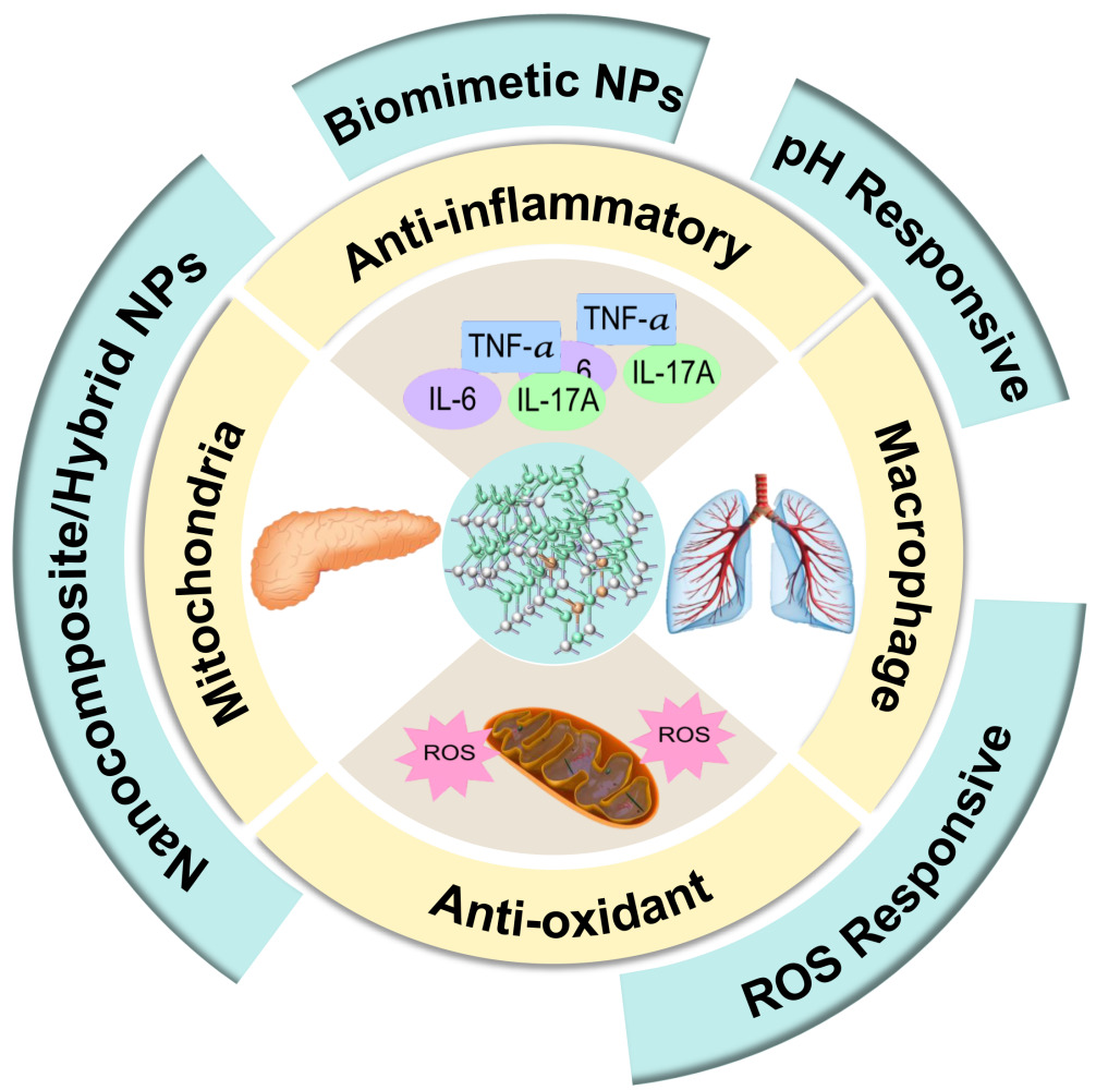
Obeng E, Ergashev A, Yao Q, Wu W, Chen G
International Journal of Nanomedicine 2025, 20:9517-9558
Published Date: 31 July 2025
Intelligent Nanomedicine Systems Utilizing Diverse Nanoparticles for Osteosarcoma Therapy: A Review
Cai J, Yang H, He Z, Wu C
International Journal of Nanomedicine 2025, 20:14115-14130
Published Date: 25 November 2025
Precision Pediatric Cancer Nanomedicine: Advancing Personalized Nano Therapies to Reduce Non-Communicable Diseases Through AI-Driven 3D-Printed Drugs
Choudhary N, Kumar D, Jyoti TP, Prajapati BG, Kchaou M, Webster TJ, Faiyazuddin M
International Journal of Nanomedicine 2026, 21:575214
Published Date: 13 February 2026
Decoy Nanoparticles and Protein Corona Modulation: A Novel Frontier in Sepsis Treatment
Denisova ER, Volkova DS, Streltsova VV, Kopytov SA, Krivosheev AS, Panfilova EV, Yan N, Kostyushev D, Ganjalikhani-Hakemi M, Pokrovsky VS, Lopus M, Golovin AV, Zamyatnin Jr AA, Celia C, Parodi A
International Journal of Nanomedicine 2026, 21:593748
Published Date: 27 April 2026