Back to Journals » International Journal of Nanomedicine » Volume 21
Nanoparticles in the Treatment of Renal Fibrosis
Authors Wu Y, Zhang W, Zhu L, Liu L
Received 30 November 2025
Accepted for publication 5 March 2026
Published 10 March 2026 Volume 2026:21 585576
DOI https://doi.org/10.2147/IJN.S585576
Checked for plagiarism Yes
Review by Single anonymous peer review
Peer reviewer comments 5
Editor who approved publication: Prof. Dr. RDK Misra
Ying Wu, Wen Zhang, Li’ou Zhu, Lili Liu
Department of Pathology, Wuhan Children’s Hospital (Wuhan Maternal and Child Healthcare Hospital), Tongji Medical College, Huazhong University of Science and Technology, Wuhan, 430016, People’s Republic of China
Correspondence: Lili Liu, Email [email protected]
Abstract: Renal fibrosis represents a key pathological hallmark of chronic kidney disease (CKD), a prevalent condition worldwide that currently lacks effective therapies. Nanotechnology offers transformative potential for renal fibrosis treatments. Engineered nanoparticles (NPs), with tunable physicochemical properties such as size, surface charge, and shape, enable targeted and controlled drug delivery. Furthermore, active targeting strategies can increase nanoparticle uptake by specific cell types or structures in kidneys. Although nanomedicine-based strategies for renal fibrosis remain at an early, predominantly preclinical stage, accumulating evidence from experimental models suggests substantial potential for improving therapeutic precision and efficacy in fibrotic kidney disease. This review integrates recent insights into the pathogenesis of renal fibrosis, NP design strategies, along with advances in NP-based therapeutics, highlighting nanomedicine as a promising approach for precise and effective intervention in CKD-associated renal fibrosis.
Keywords: renal fibrosis, chronic kidney disease, NPs, nanomedicine, drug delivery
Introduction
CKD is defined as a persistent structural or functional abnormality of the kidneys, such as, renal glomerular filtration rate (GFR) reduction (< 60 mL/min/1.73 m2) or albuminuria > 30 mg per 24 h.1 Arising from diverse etiologies, including diabetes, hypertension, and primary glomerular diseases, CKD represents a major global health concern, with a prevalence exceeding 10% and an even higher incidence in elderly populations.2 Patients with CKD frequently develop multiple complications, imposing substantial clinical and economic burdens. Renal fibrosis constitutes a critical pathological hallmark in the progression of CKD, characterized by myofibroblast proliferation and extracellular matrix (ECM) accumulation, ultimately leading to a decline in renal function.3 The severity of renal fibrosis closely correlates with disease prognosis, making it a key therapeutic target in efforts to prevent CKD progression.
Traditional systemic therapies, including sodium-glucose cotransporter 2 inhibitors, non-steroidal mineralocorticoid receptor antagonists, and glucagon-like peptide-1 receptor agonists, are often limited by poor kidney-specific targeting, short circulation half-life, low renal drug utilization, and substantial off-target toxicities.4,5 In addition, the unique anatomy of the kidney, characterized by specialized filtration barriers, pronounced cortical medullary compartmentalization, and highly selective permeability, further increases the difficulty for drugs to reach and act on specific pathological sites, resulting in suboptimal tissue exposure and therapeutic efficacy.6,7 Collectively, these challenges underscore the need for advanced drug delivery systems capable of navigating renal physiological barriers and enabling more precise therapeutic intervention. In this context, nanotechnology-based delivery platforms offer unique advantages through their tunable physicochemical properties and targeting capacity.5,8
NPs are materials engineered with atomic or molecular precision to exhibit unique physicochemical properties.9 Although classical nanoparticle definition restricts size to the nanoscale (1–100 nm), this criterion excludes many carrier systems used in biomedical applications. In the medical literature, carrier systems up to 1000 nm are frequently referred to as nanocarriers, and it has been proposed that nanocarriers be defined based on both function (ability to encapsulate or bind active ingredients) and size (at least one dimension <1000 nm).10,11 Structurally, most NPs consist of an inner core or matrix surrounded by an outer shell, providing a versatile platform for encapsulating or conjugating a wide variety of therapeutic and diagnostic agents, including small molecules, proteins, peptides, nucleic acids, and imaging probes.12 In recent years, NP-based therapeutic strategies for renal fibrosis have attracted increasing attention. A growing number of preclinical studies have explored NPs for the delivery of small-molecule drugs, nucleic acids, and biological agents aimed at modulating key fibrotic pathways, such as inflammation, myofibroblast activation, and extracellular matrix deposition.5 Although most renal nanotherapeutic approaches remain at the preclinical stage, these advances highlight the potential of nanotechnology to address long-standing challenges in the treatment of renal fibrosis.
In this review, we systematically reviewed the pathological mechanisms of renal fibrosis, the design principles of NPs for renal targeting, and presented representative examples of NP-based therapies in renal disease. In addition, we discussed the current challenges in clinical translation and provided perspectives on future directions for the development of nanomedicines targeting renal fibrosis.
Major Cell Types Participating in Renal Fibrosis
Renal fibrosis is increasingly recognized as a maladaptive extension of the normal wound-healing response following kidney injury.13,14 In the early stages, transient ECM deposition may provide structural support and limit further damage. However, upon persistent or repetitive injury, this reparative response becomes dysregulated, leading to uncontrolled ECM protein accumulation, nephron loss and vascular rarefaction.15,16 The resulting lesions, including glomerulosclerosis, tubular atrophy, interstitial fibrosis, and vascular remodeling, collectively compromise renal filtration and drive progressive renal failure.17,18
The renal tubular epithelial cells (RTECs) and myofibroblasts are the main participants in renal fibrosis. Upon injury, resident mesenchymal cells are widely recognized to activate and differentiate into myofibroblasts,19–21 which secrete pro-inflammatory mediators and ECM proteins. Instead of generating myofibroblasts,22 tubular epithelial cells promote the progression of fibrosis by acting as critical signaling hubs, mediating partial epithelial-to-mesenchymal transition (EMT),23 inflammatory responses,24 senescence,25 and intercellular crosstalk.26 In addition, other resident and infiltrating cell types, including mesangial cells, podocytes, and vascular smooth muscle cells, as well as macrophages, lymphocytes, and fibrocytes recruited from the circulation, also contribute to the renal fibrosis.27–31
Myofibroblasts
In normal adult kidneys, type IV collagen is the predominant basement membrane component.32,33 During renal interstitial fibrosis, fibrillar collagens, particularly type I and type III, are markedly upregulated, constituting the bulk of extracellular matrix deposition.34,35 This pathological ECM accumulation is largely attributed to activated myofibroblasts, which are rare in normal kidneys but markedly increased in fibrotic kidneys.36 Myofibroblasts possess characteristics of both fibroblasts and smooth muscle cells. The hallmark marker is α-smooth muscle actin (α-SMA), which forms stress fibers essential for matrix adhesion and cell contraction, facilitating matrix remodeling and wound closure.
However, the origin and heterogeneity of myofibroblasts remain incompletely understood. Resident mesenchymal cells, including fibroblasts, mesenchymal stem cells (MSCs), and pericytes, are considered common origins of myofibroblasts. Bone marrow-derived cells (macrophages and fibrocytes) and endothelial cells may also contribute to it.37–39 But these studies are still limited and additional evidence is needed to substantiate their contribution. Recent single-cell sequencing of CKD human kidneys40 identified three principal platelet-derived growth factor receptor (PDGFR)β+ myofibroblast sources: PDGFRα+ MEG3+ fibroblasts, COLEC11+ CXCL12+ fibroblasts, and PDGFRα− RGS5+ NOTCH+ pericytes. Notably, increases in ECM genes in tubular epithelial cells are minimal, suggesting a minor role for EMT in renal fibrosis. The transition of pericytes or fibroblasts to myofibroblasts is regulated by activating protein-1 (AP-1) in early stage and transforming growth factor (TGF)-β1 in late stage, with transcription factor Runx1 driving transdifferentiation and upregulating several myofibroblast genes.41
Myofibroblasts are highly responsive to TGF-β1, which is the main regulator of myofibroblast differentiation.42 Experimental evidence strongly supports the significant role of activated myofibroblasts in renal fibrosis. Deletion of αV integrin, which is critical for TGF-β1 signaling, in mouse PDGFR-β+ cells reduce fibrosis in kidney unilateral ureteral obstruction (UUO), lung, and liver fibrosis models.43 Consistently, a potent small-molecule inhibitor of αV integrin replicates this antifibrotic phenotype.44 Furthermore, specific ablation of proliferating α-SMA+ myofibroblasts via a herpes simplex viral thymidine kinase system results in a 50–60% decrease in collagen deposition and interstitial fibrosis across multiple renal fibrosis models, thereby strongly indicating that proliferating myofibroblasts are principal cells driving fibrogenesis.37
RTECs
RTECs are highly metabolically active and contain abundant mitochondria to support their reabsorptive and transport functions. Therefore, they are particularly vulnerable to various damages, such as ischemia, toxins, proteinuria, and metabolic disturbances. After injury, RTECs lose their polarity and cell-cell contacts, inhibiting normal tubular function and initiating repair processes.16 However, RTECs are both victims and active participants in renal fibrosis through complex mechanisms. When damage exceeds renal regenerative capacity, pathological changes drive RTECs to contribute to fibrosis through paracrine signaling and maladaptive cellular responses.
Inflammation is critical for tubular injury and fibrosis. Injured RTECs recruit and activate inflammatory cells, including monocytes/macrophages, lymphocytes, dendritic cells, and mast cells.45 Proinflammatory M1 macrophages release cytokines such as interleukin (IL)-6, tumor necrosis factor α, IL-12, and nitric oxide, exacerbating tubular apoptosis and dysfunction.46 Profibrotic macrophages secrete TGF-β1 to activates fibroblasts and remodels ECM.47 TGF-β1 can also induce partial EMT in RTECs, which is characterized by dedifferentiation, loss of polarity, and acquisition of some mesenchymal traits, without a full transdifferentiation. Partial EMT enables RTECs to secrete profibrotic and proinflammatory mediators that contribute to fibroblast activation and ECM production, indirectly exacerbating fibrosis.48
Apart from TGF-β1, several signaling pathways also regulate RTEC responses. Wnt/β-catenin signaling is transiently upregulated after injury to inhibit p53 and Bax expression and facilitate RTEC survival;49 however, prolonged activation induces sustained epithelial dedifferentiation and interstitial fibrosis.50 Hedgehog (Hh) ligands (Sonic and Indian Hh) show similar paracrine effects. They are increased in injured RTECs and accompanied with Hh pathway activation. Hh ligands activate interstitial pericytes and peritubular fibroblasts.51 The Notch signaling pathway involves Notch receptors (Notch1-Notch4) and their ligands, Delta and Jagged. Activation occurs through ligand–receptor interactions between neighboring cells, triggering intracellular signaling cascades that regulate gene expression critical for cell fate determination.52,53 In chronic kidney disease, Notch signaling is reactivated, especially in epithelial cells, where it drives dedifferentiation and proliferation while inhibiting differentiation.54 Notch activation is both necessary and sufficient to promote tubulointerstitial fibrosis, highlighting its key role in kidney fibrosis progression.
Design Criterion of NP Targeting Kidney
NPs can be produced from both organic and inorganic materials. Organic systems include liposomes, extracellular vesicles (EVs), biomimetic NPs, polymeric NPs, dendrimers, nanogels, and carbon-based nanostructures, while inorganic platforms encompass quantum dots, magnetic iron oxide particles, and metallic NPs.55–57
In chronic renal fibrosis research, several classes of nanoparticle delivery systems have been investigated as platforms for antifibrotic therapy. Lipid-based nanoparticles represent a major class and include liposomes and solid lipid nanoparticles, which are structurally defined by lipid bilayer or lipid-core architectures that allow incorporation of drugs within aqueous or lipid compartments.4 EVs, naturally secreted lipid bilayer vesicles, have also emerged as promising nanocarriers due to their intrinsic biocompatibility, ability to transfer proteins and nucleic acids, and potential for cell-specific targeting.58 Biomimetic organic nanoparticles, including biomimetic high-density lipoprotein (bHDL)-like nanoparticles and EV-mimetic assemblies, are distinguished by the incorporation of endogenous or biomimetic components that recapitulate structural features of natural lipoproteins or cellular vesicles.59 Polymeric nanoparticles constitute one of the most widely studied categories and are typically fabricated from biodegradable polymers such as poly(lactic-co-glycolic acid) (PLGA) and related copolymers, in which therapeutic agents are encapsulated within a degradable polymer matrix.60 Dendrimers are highly branched, monodisperse polymeric nanocarriers characterized by a well-defined architecture and multiple surface functional groups.61 In renal diseases, dendrimers have mainly been explored for acute kidney injury treatment and diagnostic detection.62,63 However, their application in renal fibrosis therapy remains relatively limited. Beyond matrix nanoparticles, amphiphilic block copolymers can self-assemble into micelles that encapsulate therapeutic agents within nanoscale cores.64 Nanogels constitute a further class of organic nanocarriers and are characterized by three-dimensional cross-linked hydrophilic polymer networks, commonly formed from materials such as poly(N-isopropylacrylamide) or chitosan derivatives.65
A core strength of nanotechnology in medicine lies in the ability to control key physical parameters to optimize bioavailability, biodistribution, and target specificity. Such flexibility in design has led to two principal renal targeting strategies (Figure 1): passive targeting, which relies on physicochemical properties like size, surface charge and shape to navigate biological transport barriers,66 and active targeting, which utilizes receptor-ligand interactions to achieve precise molecular recognition at desired tissue sites.67 Active targeting can be achieved using ligands such as antibodies, antibody fragments, peptides, proteins, aptamers, or small molecules that bind to disease-specific biomarkers.
 |
Figure 1 Nanoparticle properties to facilitate renal accumulation. Endothelial cells, glomerular basement membrane, and podocytes with slit diaphragms form the glomerular filtration barrier. Nanoparticle size, charge, shape, surface modification, and active targeting are the main factors in design to target kidney. |
NP Size
The effectiveness and biodistribution of NPs in kidney disease are critically influenced by their size. Among the various physicochemical attributes of nanomaterials, particle diameter stands out as a major determinant of filtration, retention, clearance, and hence application potential in renal pathology.68
The size of NPs is a critical determinant of their ability to traverse the glomerular filtration barrier (GFB), a key structural and functional interface governing renal clearance and kidney-specific drug delivery.68,69 The GFB acts as a sophisticated multilayered filtration system separating the bloodstream from the urinary space within the renal glomerulus. It is composed of three major layers working in concert to regulate molecular passage based on size and charge. The innermost layer, the fenestrated glomerular endothelial cells, contains slit openings approximately 60–80 nm in diameter that facilitate plasma flow while retaining larger macromolecules. The middle layer, the glomerular basement membrane, is a dense, non-cellular extracellular matrix network with an average pore size of roughly 10 nm and a predominantly negative electrostatic charge that repels anionic macromolecules. The outermost layer consists of the interdigitating foot processes of podocytes, which are separated by slit diaphragms approximately 12 nm wide and reinforced by specialized proteins such as nephrin and podocin.70 Together, these layers establish a precisely regulated permeability threshold that determines the renal filtration of NPs.
Physiological studies indicate that passive filtration across the intact GFB is governed by molecular size and charge selectivity. Small solutes traverse the barrier primarily via convective transport driven by the transcapillary hydrostatic pressure gradient, which outweighs the opposing oncotic pressure. In contrast, larger macromolecules are effectively excluded under physiological conditions due to steric hindrance and electrostatic repulsion.71,72 Most NPs with size ranging from 1–10 nm, such as quantum dots or ultrasmall gold NPs, readily pass through the GFB and are efficiently cleared via urine, leading to low renal retention.73–75 Those in the 10–20 nm range can still cross the GFB, especially if composed of soft organic materials.76,77 It was previously thought that nanoparticles needed to be smaller than approximately 50–70 nm to pass through the fenestrations of the glomerular endothelium. However, larger particles can also accumulate in the kidney. For example, 90 nm Polyethylene glycol (PEG)-PLGA nanoparticles loaded with dexamethasone acetate and 79 nm PEGylated gold nanoparticles were reported to exhibit high renal accumulation because of their association with the mesangial cells, which directly connected to the fenestrated endothelium outside the glomerulus capillaries.78,79
Theoretically, larger NPs (>100 nm) are typically excluded by the GFB. However, it has been reported that some large NPs can accumulate within proximal tubule epithelial cells. Wyss et al utilized transmission electron microscopy on kidney and observed the presence of intact spherical PLGA polymeric NPs with diameters ranging from 130 to 160 nm in glomerular vascular tuft, proximal convoluted tubule (PCT) cells and the tubule lumen.80 They proposed that those NPs were transported from the vasculature of the glomerular tuft, taken up by PCT cells and then secreted into tubular lumen. However, it remains to be proven. Williams et al synthesized polymer-based mesoscale NPs of 400 nm diameter and intravenously injected in mice.81 The accumulation of NPs in the kidney was approximately 7-fold higher than in other organs, and they were primarily localized in the basal membrane of proximal tubule epithelial cells. They proposed that those NPs were initially endocytosed by endothelial cells of the peritubular capillaries, as demonstrated in vitro and supported by previous studies. Subsequently, they were transcytosed across the endothelial cells and released into the tubulointerstitial space between the capillaries and proximal tubule epithelial cells. From this interstitial space, they were taken up by proximal tubule epithelial cells via basolateral endocytosis, as confirmed in vitro. Similarly, Deng et al also reported that PLGA-b-mPEG mesoscale NPs approximately 400 nm in diameter selectively accumulated in proximal tubule cells, exhibiting the highest fluorescence intensity in the kidney compared to other organs.82 Although these studies provide convincing evidence that some mesoscale NPs selectively target proximal tubule epithelial cells and propose plausible transport mechanisms, in vivo validation of these pathways remains limited. The exact cellular and molecular processes governing nanoparticle uptake, transcytosis, and basolateral entry into tubular cells have yet to be rigorously confirmed.
During renal fibrosis, GFB structural remodeling, including basement membrane thickening with widened pores, and podocyte detachment resulting in the disruption of slit diaphragm integrity, leads to a marked reduction in size-selective filtration capacity.8,83 Mesangial expansion and increased ECM deposition can also introduce additional barriers to diffusion.84 However, the extent to which mesangial remodeling and ECM accumulation modulate renal nanoparticle uptake has not been systematically investigated, which represents an important gap for future studies aimed at optimizing kidney-targeted nanomedicine.
NP Charge
NP charge plays an important role in determining their interaction with the kidney, particularly with the GFB, which exhibits charge selectivity due to the abundance of negatively charged heparan sulfate in the endothelial glycocalyx and anionic proteoglycans on the glomerular capillary wall and podocyte glycocalyx.85 These structural features create an electrostatic barrier that preferentially restricts the passage of negatively charged molecules, while allowing neutral and positively charged molecules to filter through.74 Consequently, NPs of different surface charges, even when similar in size, display distinct renal biodistribution and clearance profiles.
Generally, positively charged NPs traverse the GFB more efficiently than negatively charged counterparts. Experimental studies using quantum dots74 and micelles86 have consistently demonstrated that cationic NPs exhibit higher renal accumulation and faster urinary excretion compared to anionic particles of comparable size. In terms of renal clearance, positively charged NPs show markedly enhanced kidney localization compared with their negatively charged analogs. Small cationic NPs, such as amine-functionalized copper sulfide or silicon nanodots, achieve nearly complete renal clearance within 24 hours after injection.82,87 In contrast, negatively charged gold NPs of similar hydrodynamic diameters display significantly lower clearance efficiency,88,89 illustrating that surface charge can modulate renal elimination kinetics.
Nevertheless, charge-dependent renal processing is not solely governed by simple electrostatic interactions. It has been reported that negatively charged Ficoll displays increased glomerular filtration compared to neutral Ficoll.90,91 Among negatively charged nanomaterials, there is an inverse correlation between surface charge and renal clearance efficiency. Specifically, nanomaterials with more uniform and stronger negative surface charges exhibit higher renal clearance rates.88,92
Indeed, NPs with high absolute zeta potential, whether positive or negative, tend to bind serum proteins more strongly, promoting opsonization and uptake by the mononuclear phagocyte system, thereby reducing renal filtration efficiency.93,94 Conversely, NPs with near-neutral or zwitterionic surfaces had minimal macrophage uptake, which enables longer systemic circulation.73,95,96 Neutral and zwitterionic coatings, such as sulfobetaine or organosiloxane, have been shown to preserve colloidal stability in serum and facilitate kidney entry.97,98
Overall, the interplay among NP charge, surface chemistry, and protein interactions critically influences renal transport, clearance, and tissue retention. While cationic surfaces promote rapid renal filtration and excretion, neutral or zwitterionic surface modifications provide a more balanced approach, enhancing NP stability and prolonging circulation by reducing opsonization, thereby improving renal targeting and biodistribution. Importantly, the precise in vivo dynamics of NPs at the GFB remain poorly understood, underscoring the need for further investigation into renal NP transport.
NP Shape
NP geometry has been increasingly recognized as a critical factor influencing renal clearance and biodistribution. In the circulation, convective forces in blood flow dominate over Brownian motion, rendering shape a major determinant of hydrodynamic behavior and biological interactions.99 Non-spherical NPs, including rods, disks, filamentous micelles, and sheets, exhibit distinct margination dynamics and altered interactions with immune cells compared to spherical particles.100–102 NP with high aspect ratio, such as nanorods and nanoworms, often display prolonged circulation half-lives due to reduced macrophage uptake under shear flow, which in turn affects their organ distribution patterns.101,103–105
Recent studies have shown that particle aspect ratio can modulate glomerular filtration efficiency independent of molecular weight. For instance, single-walled carbon nanotubes (1.2 nm in diameter, 100–1000 nm in length) can pass through the GFB despite molecular weights (300–500 kDa) exceeding the renal cutoff for globular proteins.106–108 Their narrow cross-sectional dimension allows alignment with flow and insertion of their longitudinal axis into filtration slits, suggesting that directional orientation facilitates filtration. Comparable effects of geometry were found in polymeric dendrimers. 18 nm diameter Spherical polyamidoamine dendrimer with 2 kDa outer layer PEG showed negligible renal clearance (<10% ID/g), whereas the addition of two 20 kDa PEG chains transformed the NPs into elongated, cylindrical structures that achieved high kidney excretion (~80% ID/g).109,110
Furthermore, temporal studies indicate that geometry not only determines the extent but also the kinetics of kidney accumulation. Shorter rods and smaller anisotropic particles tend to exhibit early renal uptake, whereas longer or flexible structures show delayed but sustained accumulation due to prolonged circulation and reduced reticuloendothelial recognition.111
Beyond simple aspect ratio effects, three-dimensional geometries also modulate renal interactions and clearance behavior.112,113 DNA origami nanostructures with triangular, rectangular and tubular geometries have been reported to exhibit preferential renal accumulation in both healthy mice and mice with acute kidney injury, as evidenced by noninvasive quantitative imaging after systemic administration.114 Radiolabelled intact DNA origami constructs localized prominently in the kidneys with comparatively low hepatic uptake, whereas partially folded DNA origami assemblies and the scaffold M13 single‑stranded DNA exhibited predominant liver sequestration, indicating a marked impact of structural integrity on biodistribution. PET imaging and ex vivo analyses demonstrated that all three fully folded DNA origami architectures accumulated in kidney tissue significantly more than shorter single‑stranded or partially folded analogues, underscoring the role of proper folding and compact architecture in modulating organ partitioning. DNA tetrahedron nanoparticles, a minimal wireframe DNA geometry, also displayed efficient renal clearance in mouse models, further illustrating that compact DNA nanostructures can be efficiently processed by the renal system.76 Moreover, kidney-targeted DNA tetrahedral frameworks have been exploited for targeted cytosolic siRNA delivery, showing pronounced renal uptake and functional delivery to tubular cells.115
Taken together, these studies highlight that NP shape exerts a multifactorial influence on renal pharmacokinetics. Both simple anisotropy, such as high aspect ratio rods and cylinders, and more complex three-dimensional architectures, such as DNA nanostructures, can modulate the kinetics and tissue-specific distribution of nanoparticles.
NP Surface Modification
Various proteins in biological fluids can adsorb onto the NP surface forming protein corona. Protein corona not only changes the NP physicochemical properties, including charge, hydrophobicity, and functional ligands but also imparts a new biological identity that dictates recognition by the mononuclear phagocyte system (MPS).116 Surface engineering strategies have been widely employed to overcome the limitation. PEG conjugation remains a well-established approach to sterically hinder protein adsorption and reduce MPS uptake, thereby prolonging systemic circulation.117 However, PEGylation can also reduce cellular uptake by targeting cells,118,119 highlighting the need to balance circulation longevity with effective delivery.
Beyond PEGylation, additional surface functionalization strategies, including short amino acid repeat peptides, single amino acid modifications, and prodrug-like display, have emerged to achieve more precise targeting. L-serine modified polyamidoamine (PAMAM) dendrimers have been reported to preferentially accumulate in the proximal tubules of the renal cortex after intravenous administration in mice.120,121 In these constructs, multiple serine residues are covalently attached to the PAMAM surface. Because serine is an endogenous molecule with well-defined physicochemical characteristics, this modification has been suggested to provide advantages over conventional kidney-targeting moieties, including safety and synthetic simplicity.122 Chen et al engineered a multifunctional kidney-targeted nanoplatform based on a PAMAM dendrimer.123 In this system, dendrimers were modified with multiple glutathione (GSH) moieties and covalently linked to triptolide (TP) to form a prodrug-type dendrimer–drug conjugate. The design exploited the high expression of γ-glutamyltransferase (GGT) in glomerular capillary endothelial cells of childhood nephrotic syndrome. GGT catalyzed γ-glutamyl transfer reactions of GSH, generating positively charged primary amine groups on the carrier surface. This charge conversion facilitated rapid caveolae-mediated endocytosis and subsequent transcytosis, thereby enhancing renal accumulation and cellular uptake. TP was progressively released through intracellular enzymatic hydrolysis, providing sustained drug exposure.
NP Active Targeting
Beyond the passive accumulation determined by NP physicochemical parameters such as size, charge, and shape, active targeting strategies have emerged as an essential approach to achieve precise renal drug delivery.
In the glomerular compartment, mesangial cells express a variety of receptors that can be leveraged for targeted nanotherapy, including mannose receptors, α8 integrin, Thy1.1 antigen, and αvβ3 integrin.124–126 NPs functionalized with ligands recognizing these receptors have shown enhanced accumulation and therapeutic efficacy. For instance, mannose-modified polycationic cyclodextrin NPs have been successfully used to deliver siRNA selectively to mesangial cells.127 α8 integrin-targeted liposome-PLGA NPs carrying dexamethasone and captopril significantly ameliorated glomerular inflammation and fibrosis.128 And Thy1.1-directed liposomes and dual-ligand systems targeting both AT1r and αvβ3 integrin have demonstrated precise mesangial localization and suppression of extracellular matrix deposition.129,130 In addition, peptide sequence CSAVPLC targeting glomerular endothelium successfully increases kidney accumulation of its conjugated NP by 2.6 times.131 Albumin-conjugated NPs target glucocorticoid receptors and the neonatal Fc receptor, which is highly expressed on podocytes, to enhance localization and therapeutic efficiency in glomeruli.132,133
In renal tubules, active targeting primarily relies on the recognition of receptors expressed on RTECs, notably megalin and cubilin, which mediate endocytic uptake of various ligands.134,135 Nanocarriers conjugated with aminoglycoside antibiotics gentamicin and neomycin accumulate predominantly in kidney through megalin receptor binding, resulting in a 13-fold enhancement in gene transfection efficiency compared with unmodified NPs.136 Similarly, polymyxin B or low molecular weight chitosan (LMWC), which have also shown strong affinity for megalin, promotes NP uptake into proximal tubular cells and supports the delivery of therapeutic cargos such as metformin or fluorescent reporters.137,138 Additionally, the kidney-targeting peptide (KKEEE)3K has been identified as a specific binding ligand for megalin on proximal tubular cells.139 When conjugated to 15 nm PEGylated micelles, this peptide enabled selective renal accumulation.140 These designs highlight the potential of megalin-mediated endocytosis for treating tubular fibrosis and other chronic kidney pathologies.
Emerging active-targeting strategies also take advantage of pathological microenvironments. In fibrotic kidneys, ligand modification of NPs with kidney injury molecule-1 (KIM-1) antibodies allows selective binding to injured tubular epithelial cells, markedly increasing local drug concentration and therapeutic benefit.141,142 Furthermore, environment-responsive systems, such as pH-sensitive NPs that disassemble under acidic conditions, enable controlled drug release while minimizing systemic exposure.143
In addition to synthetic nanoparticle platforms, biologically derived vesicles such as exosomes have emerged as a distinct class of actively targeted nanocarriers for renal delivery.144 They have more abundant entry routes than artificial nanoparticles, including receptor-mediated endocytosis, phagocytosis, macropinocytosis, lipid raft interactions, and direct fusion.145–147 Exosomes can be engineered via surface modification or cargo loading to increase renal uptake and targeting efficiency.148 Endogenous loading integrates therapeutic molecules into exosomes through genetic modification of parent cells,149 while exogenous loading introduces small molecules or RNAs into pre-isolated exosomes using chemical or physical methods.150
Taken together, these active targeting strategies underscore the transition from nonspecific passive accumulation to precision-guided renal nanomedicine. The integration of peptides, antibodies, and stimuli-responsive designs provides multiple options for achieving high-efficiency, site-selective NP delivery to distinct kidney compartments.
NPs in the Treatment of Renal Fibrosis
Targeting RTECs
There are many ongoing studies focusing on NPs targeting RTECs to treat renal fibrosis (Table 1). These studies include various strategies, such as assembling NPs with antibodies against KIM-1, which is expressed in injured RTECs, and utilizing chitosan-based materials to achieve effective targeting.
 |
Table 1 NPs Targeting RTECs in the Treatment of Renal Fibrosis |
In chronic kidney disease, persistent endoplasmic reticulum dysfunction and stress-driven apoptosis accelerate renal damage.160 Thapsigargin (TG) induces moderate endoplasmic reticulum stress and activates autophagy to protect renal cells from oxidative injury.161 Building on this mechanism, KIM-1-targeted TG NPs (KIM-1-TG NPs) were developed to achieve kidney-specific delivery through targeting injured RTECs. These NPs were made from PLGA, and remained stable at neutral pH but release TG within acidic intracellular environments, ensuring precise, localized drug action. In an adenine diet-induced CKD mouse model, low doses (0.2 mg/kg) of KIM-1-TG NPs effectively reduced renal injury.151 Lin et al142 utilized similar carrier to synthesized KIM-1 targeting NPs loaded with resveratrol (Res). Res is a natural polyphenol exhibiting beneficial effects in several renal diseases, such as diabetic nephropathy, drug-induced injury, and unilateral ureteral obstruction.162–164 In adenine -induced CKD mouse model, Res NPs increased AMPK and inhibited the Akt/mTOR signaling pathways to induce autophagy, therefore suppressing NLRP3 inflammasome and ameliorating CKD. It is reported that KIM-1-bHDL NPs loaded with both anti-inflammatory drug TP and anti-fibrotic drug nintedanib (BIBF) reduced renal fibrosis in UUO model, folic acid model and adenine model mice via remodeling the fibrosis niches.152 Exosomes could also be engineered as targeted delivery vehicles to injured renal tubular epithelial cells. For example, red blood cell–derived extracellular vesicles (REVs) have been modified with a KIM-1-binding peptide, enabling selective accumulation in injured tubules. Using this platform, siRNAs against the transcription factors P65 and Snai1 were efficiently delivered to ischemic kidneys, resulting in reduced inflammation and fibrosis.153
LMWC can be selectively taken up by RTECs through megalin-mediated endocytosis.165 Therefore, LMWC and its modified derivatives have been widely employed in the treatment of renal fibrosis. Wischnjow et al154 synthesized GFP-loaded metformin-grafted chitosan NPs (CS-MET/GFP NPs). After intravenous injection, those NPs accumulated in kidney and released inside the cells, exerting anti-apoptotic, anti-inflammatory, and anti-fibrotic effects in UUO mouse model. Ghavimishamekh et al155 also reported that oral intake of insulin-loaded trimethyl chitosan NP reduced TGF-β1 expression and amelibrated diabete-induced nephropathy in type 1 diabetic rat model. Qiao et al developed a kidney-specific drug nanocomplex using coordination bonding with catechol-modified low molecular weight chitosan (HCA-Chi) as the polymeric carrier.156 The stable nanocomplex circulates at physiological pH, while upon lysosomal internalization, acidic pH triggers partial drug release by breaking coordination bonds, allowing subsequent transmembrane transport and paracrine effects in the tubulointerstitium. Emodin is a natural anthraquinone compound that exerts its anti-renal fibrosis effects via multiple mechanisms, including mediating mitochondrial homeostasis,166 inhibiting TGF-β1/Smad signaling pathway,167 and regulating autophagy168 and apoptosis.169 The HCA-Chi-Zn-emodin ternary nanocomplex effectively attenuates renal fibrosis by targeting and suppressing myofibroblast activation, as demonstrated by reduced α-SMA expression in UUO-injured kidneys. Histological analyses confirmed that this nanocomplex decreases tubular dilation, epithelial atrophy, and collagen I accumulation. In addition, HCA-Chi-Ca-salvianolic acid B (Sal B) NPs showed similar anti-fibrosis effects in UUO mouse model.157
MicroRNAs (miRNAs) are negative regulators of gene expression, through repressing mRNA translation or promoting mRNA degradation. Recent research has revealed their significant role in the progression of kidney fibrosis.170,171 However, miRNAs and siRNAs are inherently unstable and vulnerable to degradation by nucleases in biological environments. To overcome this limitation, NPs have been employed to encapsulate these nucleic acids, protecting them from nuclease-mediated degradation. miRNA-21 promoted renal fibrosis through TGF-β1/Smad3 and extracellular signal-regulated kinases/mitogen-activated protein (ERK/MAP) kinase signaling pathway.172 Geng et al138 synthesized small-sized cationic miRNA-21 inhibitor (miRi)-PCNPs which utilized LMWC-modified PLGA to specifically deliver miRNA-21 inhibitor to RTECs. In UUO model, mice treated with miRi significantly reduced extracellular matrix accumulation, while fibrosis area after miRi-PCNPs treatment was 2.5-fold lower than miRi treatment. Khaja et al158 reported that they utilized galactosamine-conjugated sterically stabilized phospholipid NPs (SSLNPs) to delivery small interfering RNA (siRNA) for connective tissue growth factor (CTGF), which was the important regulator of fibrosis in both hepatic and renal cells.173,174 Bio-distribution and pharmacokinetic studies in mice confirmed their targeted delivery to liver and kidney tissues. However, their therapeutic effect on renal fibrosis still requires further investigation.
CD44 is expressed on both glomerular and tubular cells in the kidney, including renal epithelial cells, mesangial cells, and fibroblasts.175–177 Chitosan/hyaluronan NPs carrying plasmid DNA (CS/HApDNA NPs) targets glomerular and tubular cells through HA binding CD44 receptors. CS/HApDNA NPs expressing bone morphogenetic protein 7 (BMP7) reversed renal fibrosis and regenerated tubules, whereas delivery of hepatocyte growth factor NK1 (HGF/NK1) eliminated collagen fiber deposition.159
Targeting Glomeruli and Other Cells
Beyond renal tubular epithelial cells, NP therapies have been engineered to target other renal cell populations for precise and effective treatment (Table 2). Zhou et al128 utilized PEG-modified liposome-PLGA, α8 integrin antibodies, dexamethasone (DXMS) and captopril (CAP) to synthesize DXMS/CAP@PLGA-ILs. The resulting NPs have a core-shell structure, suitable particle size (~119 nm), low cytotoxicity, and the ability to specifically accumulate in the glomerular mesangial cell region. DXMS/CAP@PLGA-ILs were intravenously injected in a mouse model of mesangial proliferative glomerulonephritis (MesPGN). In vivo pharmacodynamic studies demonstrated that DXMS/CAP@PLGA-ILs significantly decreased the levels of inflammatory factors, fibrotic markers, and reactive oxygen species (ROS), thereby alleviating renal inflammation and fibrosis. The design of 2i@DuaLR NPs mainly utilized passive targeting strategy: the size of 2i@DuaLR is 110 nm, which was larger than podocytes feet slits and smaller than glomerular endothelium pores; cationic octa-arginine (R8) peptide in 2i@DuaLR NPs was trapped in glomeruli with polyanionic surface glycoprotein.178 2i@DuaLR NPs, which were loaded with both p38α MAPK and p65 siRNA, successfully silenced p38α MAPK and p65 genes in mesangial cells and endothelial cells, and eventually alleviated proteinuria, inflammation and excessive extracellular matrix deposition.
 |
Table 2 NPs Targeting Glomeruli and Other Cells in the Treatment of Renal Fibrosis |
Rosiglitazone (RSG) is an exogenous agonist of peroxisome proliferator-activated receptor (PPAR)γ, which is widely expressed in the medullary collecting duct, glomeruli, and proximal tubular cells.185 RSG-induced PPARγ activation inhibits inflammatory cytokine secretion and prevents ECM synthesis through inhibiting TGF-β1 signaling pathway.186 PLNPs-RSG-MBs contain ultrasound-targeted microbubbles. When exposed to ultrasound, microbubbles mediate sonoporation, which results in temporary increase of cellular membrane or microvasculature permeabilization, and promotes the intracellular RSG uptake without inducing glomerular injury.187 In the UUO rat model, PLNPs-RSG-MBs treatment with ultrasound exposure significantly reduced the expressions of TGF-β1, α-SMA and Collagen I.179
Even though chitosan is considered a powerful RTEC-targeting strategy, it was also demonstrated that chitosan carrying cyclooxygenase type 2 (COX-2) siRNA accumulated in macrophages in the obstructed kidney of UUO mice.180
Cationic bovine serum albumin (cBSA)/ miR-29b NPs loaded into shear-thinning F127-HA hydrogel (gel) could form a supramolecular hydrogel hybrid platform. A single injection of the hydrogel loaded with cBSA/miR-29b in UUO mice significantly downregulated proteins and genes related to fibrosis without affecting the normal liver or kidney functions.181
Wang et al182 reported that they engineered a delivery system for miR-29 by fusing the exosomal membrane protein Lamp2b with the rabies viral glycoprotein (RVG) peptide, which enables targeting to tissues expressing acetylcholine receptors. These engineered exosomes were administered intramuscularly in a UUO mouse model and partially attenuated renal fibrosis, which was associated with suppression of YY1 and TGF-β signaling pathways. EVs derived from genetically engineered MSCs were used as a delivery platform for erythropoietin (EPO) in the treatment of renal anemia.183 Following intraperitoneal administration in a mouse model of chronic kidney disease, EPO(+)-EVs significantly increased hemoglobin levels and improved renal function. Although the precise mechanisms governing the distribution of EVs to injured kidneys were not fully defined, the authors suggested that surface adhesion molecules and integrins on EV membranes may contribute to their accumulation in damaged renal tissue. In another study, bioactive scaffolds incorporating multifunctional EVs (mEVs) were developed to promote kidney regeneration.184 The mEVs consisted of a combination of EVs derived from SDF-1α overexpressing tonsil-derived MSCs and EVs from intermediate mesoderm cells differentiating toward kidney progenitor cells, aiming to enhance stem cell recruitment and renal lineage differentiation. These vesicles were embedded in porous PLGA-based scaffolds together with the antioxidant edaravone (EDV), which supported tubular regeneration and improved renal function through activation of the GDNF/RET signaling pathway. This work illustrates that EVs can also be integrated into scaffolds to provide sustained release and synergize with regenerative cues for kidney tissue engineering.
Challenges and Future Perspectives
Clinically, these NP-based strategies have the potential to shift renal fibrosis management from symptomatic treatment toward precise, mechanism-based interventions that can slow disease progression, improve kidney function, and reduce the need for dialysis or transplantation.
However, significant challenges remain before these approaches can be successfully translated into clinical practice. One of the primary barriers lies in the precise control of NP physicochemical properties, which critically determine renal accumulation and cellular uptake. Oversized particles may fail to penetrate the dense ECM of fibrotic kidneys, while smaller ones are prone to rapid renal clearance. Variability among patients in CKD stages introduces further complexity, as the structural and functional alterations in kidney, such as changes in filtration barrier integrity and pore size distribution, can significantly affect NP trafficking and retention.
In addition, ensuring NP stability and biocompatibility in vivo remains a formidable task. Synthetic nanocarriers, including polymeric and inorganic systems, may exhibit uncontrolled biodistribution or induce immune activation, compromising long-term safety.188,189 Although biomimetic and cell membrane-coated NPs have shown improved circulation time and immune evasion, their production still faces challenges related to scalability, standardization, and compositional heterogeneity. The absence of unified manufacturing standards and good manufacturing practice (GMP)-grade synthesis for complex nanocarriers further limits reproducibility and comparability across studies.190 Ongoing efforts toward modular, automated GMP-compatible synthesis and AI-assisted quality control are expected to bridge this gap and accelerate clinical readiness.
Another challenge arises from the limitations of current animal models, which fail to fully recapitulate human renal fibrosis. Animal models remain indispensable for evaluating the pharmacokinetics, biodistribution, and therapeutic efficacy of NP-based systems in renal fibrosis. Several studies have demonstrated cross-species correlations in NP pharmacokinetics, suggesting that, under certain conditions, preclinical models can partially predict human outcomes.191–193 However, the anatomical and histological complexity of the human kidney, along with disease-specific heterogeneity such as glomerular sclerosis and interstitial remodeling, is difficult to fully recapitulate in commonly used animal models such as UUO rodents.194 Recently, the emergence of human kidney organoids, precision-cut kidney slices, and humanized mouse models offers promising platforms to bridge this gap by providing physiologically relevant systems under human-like conditions.195–197
Looking forward, several directions may accelerate progress in this field. The integration of artificial intelligence and machine learning will optimize the nanocarrier design by predicting the ideal physicochemical parameters for renal delivery, modeling NP-ECM interaction, and guiding ligand selection based on single-cell transcriptomic data.198,199 Multi-functional and stimuli-responsive nanocarriers capable of co-delivering anti-fibrotic, anti-inflammatory, and ECM-degrading agents might be an option to overcome the multifactorial nature of renal fibrosis. Combining such systems with microfluidic synthesis and high-throughput screening technologies allow the controlled production of NPs with narrow size distributions and tunable surface chemistry suitable for clinical translation.200,201 With continued innovation in material science, advanced modeling, and humanized disease systems, NP-based therapeutics hold great promise to transform the current therapeutic landscape of renal fibrosis, shifting it from symptomatic management toward precise, mechanism-based intervention and regeneration.
Funding
This work was supported by Wuhan Municipal Health Commission (WZ22Q06).
Disclosure
The authors report no conflicts of interest in this work.
References
1. Stevens PE, Ahmed SB, Carrero JJ, et al. KDIGO 2024 clinical practice guideline for the evaluation and management of chronic kidney disease. Kidney Int. 2024;105(4S):S117–19. doi:10.1016/j.kint.2023.10.018
2. Luo S, Grams ME. Epidemiology research to foster improvement in chronic kidney disease care. Kidney Int. 2020;97(3):477–486. doi:10.1016/j.kint.2019.11.010
3. Miguel V, Shaw IW, Kramann R. Metabolism at the crossroads of inflammation and fibrosis in chronic kidney disease. Nat Rev Nephrol. 2025;21(1):39–56. doi:10.1038/s41581-024-00889-z
4. Wang Z, Zhang C. Nanomaterials for targeted therapy of kidney diseases: strategies and advances. Mater Today Bio. 2025;31:101534. doi:10.1016/j.mtbio.2025.101534
5. Yao P, Zheng Y, Li C. Precision nanotherapeutics for kidney disease: targeting inflammation and maladaptive repair. Int Urol Nephrol. 2025;58:949–965. doi:10.1007/s11255-025-04714-9
6. Schoales Z, Ghosh P, Vasylaki A, Jaimes EA, Williams R. Pathways to translation for nanomedicine in nephrology. Clin Kidney J. 2025;18(9):sfaf192. doi:10.1093/ckj/sfaf192
7. Sabiu G, Kasinath V, Jung S, Li X, Tsokos GC, Abdi R. Targeted nanotherapy for kidney diseases: a comprehensive review. Nephrol Dial Transplant. 2023;38(6):1385–1396. doi:10.1093/ndt/gfac233
8. Huang Y, Ning X, Ahrari S, et al. Physiological principles underlying the kidney targeting of renal nanomedicines. Nat Rev Nephrol. 2024;20(6):354–370. doi:10.1038/s41581-024-00819-z
9. Farokhzad OC, Langer R. Impact of nanotechnology on drug delivery. ACS Nano. 2009;3(1):16–20. doi:10.1021/nn900002m
10. Chariou PL, Ortega-Rivera OA, Steinmetz NF. Nanocarriers for the delivery of medical, veterinary, and agricultural active ingredients. ACS Nano. 2020;14(3):2678–2701. doi:10.1021/acsnano.0c00173
11. Dri DA, Rinaldi F, Carafa M, Marianecci C. Nanomedicines and nanocarriers in clinical trials: surfing through regulatory requirements and physico-chemical critical quality attributes. Drug Deliv Transl Res. 2023;13(3):757–769. doi:10.1007/s13346-022-01262-y
12. Mitchell MJ, Billingsley MM, Haley RM, Wechsler ME, Peppas NA, Langer R. Engineering precision nanoparticles for drug delivery. Nat Rev Drug Discov. 2021;20(2):101–124. doi:10.1038/s41573-020-0090-8
13. Di X, Li Y, Wei J, Li T, Liao B. Targeting fibrosis: from molecular mechanisms to advanced therapies. Adv Sci. 2025;12(3):e2410416. doi:10.1002/advs.202410416
14. Boor P, Ostendorf T, Floege J. Renal fibrosis: novel insights into mechanisms and therapeutic targets. Nat Rev Nephrol. 2010;6(11):643–656. doi:10.1038/nrneph.2010.120
15. Urban ML, Manenti L, Vaglio A. Fibrosis--a common pathway to organ injury and failure. N Engl J Med. 2015;373(1):95–96. doi:10.1056/NEJMc1504848
16. Ruiz-Ortega M, Rayego-Mateos S, Lamas S, Ortiz A, Rodrigues-Diez RR. Targeting the progression of chronic kidney disease. Nat Rev Nephrol. 2020;16(5):269–288. doi:10.1038/s41581-019-0248-y
17. Huang R, Fu P, Ma L. Kidney fibrosis: from mechanisms to therapeutic medicines. Signal Transduct Target Ther. 2023;8(1):129. doi:10.1038/s41392-023-01379-7
18. Chen TK, Knicely DH, Grams ME. Chronic kidney disease diagnosis and management: a review. JAMA. 2019;322(13):1294–1304. doi:10.1001/jama.2019.14745
19. Lin S-L, Kisseleva T, Brenner DA, Duffield JS. Pericytes and perivascular fibroblasts are the primary source of collagen-producing cells in obstructive fibrosis of the kidney. Am J Pathol. 2008;173(6):1617–1627. doi:10.2353/ajpath.2008.080433
20. Humphreys BD, Lin S-L, Kobayashi A, et al. Fate tracing reveals the pericyte and not epithelial origin of myofibroblasts in kidney fibrosis. Am J Pathol. 2010;176(1):85–97. doi:10.2353/ajpath.2010.090517
21. Picard N, Baum O, Vogetseder A, Kaissling B, Le Hir M. Origin of renal myofibroblasts in the model of unilateral ureter obstruction in the rat. Histochem Cell Biol. 2008;130(1):141–155. doi:10.1007/s00418-008-0433-8
22. Grgic I, Duffield JS, Humphreys BD. The origin of interstitial myofibroblasts in chronic kidney disease. Pediatr Nephrol. 2012;27(2):183–193. doi:10.1007/s00467-011-1772-6
23. Shi Y, Tao M, Chen H, et al. Ubiquitin-specific protease 11 promotes partial epithelial-to-mesenchymal transition by deubiquitinating the epidermal growth factor receptor during kidney fibrosis. Kidney Int. 2023;103(3):544–564. doi:10.1016/j.kint.2022.11.027
24. Sheng L, Zhuang S. New insights into the role and mechanism of partial epithelial-mesenchymal transition in kidney fibrosis. Front Physiol. 2020;11:569322. doi:10.3389/fphys.2020.569322
25. Zhang J-Q, Li -Y-Y, Zhang X-Y, et al. Cellular senescence of renal tubular epithelial cells in renal fibrosis. Front Endocrinol. 2023;14:1085605. doi:10.3389/fendo.2023.1085605
26. Guo C, Cui Y, Jiao M, et al. Crosstalk between proximal tubular epithelial cells and other interstitial cells in tubulointerstitial fibrosis after renal injury. Front Endocrinol. 2024;14:1256375. doi:10.3389/fendo.2023.1256375
27. Hu S, Hang X, Wei Y, Wang H, Zhang L, Zhao L. Crosstalk among podocytes, glomerular endothelial cells and mesangial cells in diabetic kidney disease: an updated review. Cell Commun Signal. 2024;22(1):136. doi:10.1186/s12964-024-01502-3
28. Jha JC, Dai A, Holterman CE, et al. Endothelial or vascular smooth muscle cell-specific expression of human NOX5 exacerbates renal inflammation, fibrosis and albuminuria in the Akita mouse. Diabetologia. 2019;62(9):1712–1726. doi:10.1007/s00125-019-4924-z
29. Tang PM-K, Nikolic-Paterson DJ, Lan H-Y. Macrophages: versatile players in renal inflammation and fibrosis. Nat Rev Nephrol. 2019;15(3):144–158. doi:10.1038/s41581-019-0110-2
30. Zhou Y, Luo Z, Liao C, et al. MHC class II in renal tubules plays an essential role in renal fibrosis. Cell Mol Immunol. 2021;18(11):2530–2540. doi:10.1038/s41423-021-00763-z
31. Yan J, Zhang Z, Jia L, Wang Y. Role of bone marrow-derived fibroblasts in renal fibrosis. Front Physiol. 2016;7:61. doi:10.3389/fphys.2016.00061
32. Rende U, Ahn SB, Adhikari S, et al. Deciphering the kidney matrisome: identification and quantification of renal extracellular matrix proteins in healthy mice. Int J Mol Sci. 2023;24(3):2827. doi:10.3390/ijms24032827
33. Alexakis C, Maxwell P, Bou-Gharios G. Organ-specific collagen expression: implications for renal disease. Nephron Exp Nephrol. 2005;102(3–4):e71–5. doi:10.1159/000089684
34. Baues M, Klinkhammer BM, Ehling J, et al. A collagen-binding protein enables molecular imaging of kidney fibrosis in vivo. Kidney Int. 2020;97(3):609–614. doi:10.1016/j.kint.2019.08.029
35. Magalhaes P, Pejchinovski M, Markoska K, et al. Association of kidney fibrosis with urinary peptides: a path towards non-invasive liquid biopsies? Sci Rep. 2017;7(1):16915. doi:10.1038/s41598-017-17083-w
36. Kramann R, Dirocco DP, Maarouf OH, Humphreys BD. Matrix producing cells in chronic kidney disease: origin, regulation, and activation. Curr Pathobiol Rep. 2013;1(4):301–311. doi:10.1007/s40139-013-0026-7
37. LeBleu VS, Taduri G, O’Connell J, et al. Origin and function of myofibroblasts in kidney fibrosis. Nat Med. 2013;19(8):1047–1053. doi:10.1038/nm.3218
38. Zeisberg EM, Potenta SE, Sugimoto H, Zeisberg M, Kalluri R. Fibroblasts in kidney fibrosis emerge via endothelial-to-mesenchymal transition. J Am Soc Nephrol. 2008;19(12):2282–2287. doi:10.1681/ASN.2008050513
39. Phua YL, Martel N, Pennisi DJ, Little MH, Wilkinson L. Distinct sites of renal fibrosis in Crim1 mutant mice arise from multiple cellular origins. J Pathol. 2013;229(5):685–696. doi:10.1002/path.4155
40. Kuppe C, Ibrahim MM, Kranz J, et al. Decoding myofibroblast origins in human kidney fibrosis. Nature. 2021;589(7841):281–286. doi:10.1038/s41586-020-2941-1
41. Li Z, Kuppe C, Ziegler S, et al. Chromatin-accessibility estimation from single-cell ATAC-seq data with scOpen. Nat Commun. 2021;12(1):6386. doi:10.1038/s41467-021-26530-2
42. Hoyles RK, Derrett-Smith EC, Khan K, et al. An essential role for resident fibroblasts in experimental lung fibrosis is defined by lineage-specific deletion of high-affinity type II transforming growth factor β receptor. Am J Respir Crit Care Med. 2011;183(2):249–261. doi:10.1164/rccm.201002-0279OC
43. Henderson NC, Arnold TD, Katamura Y, et al. Targeting of αv integrin identifies a core molecular pathway that regulates fibrosis in several organs. Nat Med. 2013;19(12):1617–1624. doi:10.1038/nm.3282
44. Chang Y, Lau WL, Jo H, et al. Pharmacologic blockade of αvβ1 integrin ameliorates renal failure and fibrosis in vivo. J Am Soc Nephrol. 2017;28(7):1998–2005. doi:10.1681/ASN.2015050585
45. Chung ACK, Lan HY. Chemokines in renal injury. J Am Soc Nephrol. 2011;22(5):802–809. doi:10.1681/ASN.2010050510
46. Huen SC, Cantley LG. Macrophages in renal injury and repair. Annu Rev Physiol. 2017;79(1):449–469. doi:10.1146/annurev-physiol-022516-034219
47. Wynn TA, Vannella KM. Macrophages in tissue repair, regeneration, and fibrosis. Immunity. 2016;44(3):450–462. doi:10.1016/j.immuni.2016.02.015
48. Bao Y, Shan Q, Lu K, et al. Renal tubular epithelial cell quality control mechanisms as therapeutic targets in renal fibrosis. J Pharm Anal. 2024;14(8):100933. doi:10.1016/j.jpha.2024.01.001
49. Zhou D, Li Y, Lin L, Zhou L, Igarashi P, Liu Y. Tubule-specific ablation of endogenous β-catenin aggravates acute kidney injury in mice. Kidney Int. 2012;82(5):537–547. doi:10.1038/ki.2012.173
50. Zhou D, Tan RJ, Zhou L, Li Y, Liu Y. Kidney tubular β-catenin signaling controls interstitial fibroblast fate via epithelial-mesenchymal communication. Sci Rep. 2013;3(1):1878. doi:10.1038/srep01878
51. Fabian SL, Penchev RR, St-Jacques B, et al. Hedgehog-Gli pathway activation during kidney fibrosis. Am J Pathol. 2012;180(4):1441–1453. doi:10.1016/j.ajpath.2011.12.039
52. Sharma S, Sirin Y, Susztak K. The story of Notch and chronic kidney disease. Curr Opin Nephrol Hypertens. 2011;20(1):56–61. doi:10.1097/MNH.0b013e3283414c88
53. Sweetwyne MT, Tao J, Susztak K. Kick it up a notch: notch signaling and kidney fibrosis. Kidney Int. 2014;4(1):91–96. doi:10.1038/kisup.2014.17
54. Bielesz B, Sirin Y, Si H, et al. Epithelial Notch signaling regulates interstitial fibrosis development in the kidneys of mice and humans. J Clin Invest. 2010;120(11):4040–4054. doi:10.1172/JCI43025
55. Eftekhari A, Kryschi C, Pamies D, et al. Natural and synthetic nanovectors for cancer therapy. Nanotheranostics. 2023;7(3):236–257. doi:10.7150/ntno.77564
56. Inci I, Eksin E, Buyukoz M, Yilmaz M. A review of nanoparticles in bioinks. Nanotechnology. 2025;36(34):342002. doi:10.1088/1361-6528/adf859
57. Unnikrishnan G, Joy A, Megha M, Kolanthai E, Senthilkumar M. Exploration of inorganic nanoparticles for revolutionary drug delivery applications: a critical review. Discov Nano. 2023;18(1):157. doi:10.1007/s40139-013-0026-7
58. Jing H, Tang S, Lin S, Liao M, Chen H, Zhou J. The role of extracellular vesicles in renal fibrosis. Cell Death Dis. 2019;10(5):367. doi:10.1038/s41419-019-1605-2
59. Agger EM. Novel adjuvant formulations for delivery of anti-tuberculosis vaccine candidates. Adv Drug Deliv Rev. 2016;102:73–82. doi:10.1016/j.addr.2015.11.012
60. Danhier F, Ansorena E, Silva JM, Coco R, Le Breton A, Preat V. PLGA-based nanoparticles: an overview of biomedical applications. J Control Release. 2012;161(2):505–522. doi:10.1016/j.jconrel.2012.01.043
61. Shang S, Li X, Wang H, et al. Targeted therapy of kidney disease with nanoparticle drug delivery materials. Bioact Mater. 2024;37:206–221. doi:10.1016/j.bioactmat.2024.03.014
62. Katsumi H, Takashima R, Suzuki H, et al. S-nitrosylated l -serine-modified dendrimer as a kidney-targeting nitric oxide donor for prevention of renal ischaemia/reperfusion injury. Free Radic Res. 2020;54(11–12):841–847. doi:10.1080/10715762.2019.1697437
63. Kobayashi H, Jo S-K, Kawamoto S, et al. Polyamine dendrimer-based MRI contrast agents for functional kidney imaging to diagnose acute renal failure. J Magn Reson Imaging. 2004;20(3):512–518. doi:10.1002/jmri.20147
64. Kaur J, Mishra V, Singh SK, et al. Harnessing amphiphilic polymeric micelles for diagnostic and therapeutic applications: breakthroughs and bottlenecks. J Control Release. 2021;334:64–95. doi:10.1016/j.jconrel.2021.04.014
65. S F, Umashankar MS, Narayanasamy D. A comprehensive review of nanogel-based drug delivery systems. Cureus. 2024;16(9):e68633. doi:10.7759/cureus.68633
66. Villaverde G, Baeza A. Targeting strategies for improving the efficacy of nanomedicine in oncology. Beilstein J Nanotechnol. 2019;10:168–181. doi:10.3762/bjnano.10.16
67. Sanna V, Sechi M. Therapeutic potential of targeted nanoparticles and perspective on nanotherapies. ACS Med Chem Lett. 2020;11(6):1069–1073. doi:10.1021/acsmedchemlett.0c00075
68. Daehn IS, Duffield JS. The glomerular filtration barrier: a structural target for novel kidney therapies. Nat Rev Drug Discov. 2021;20(10):770–788. doi:10.1038/s41573-021-00242-0
69. Haraldsson B, Nystrom J, Deen WM. Properties of the glomerular barrier and mechanisms of proteinuria. Physiol Rev. 2008;88(2):451–487. doi:10.1152/physrev.00055.2006
70. Liu M, Li X, Gao Y, et al. Breaking the fibrotic code: nanotechnology-driven advances in renal fibrosis therapy. Biomaterials. 2026;327:123738. doi:10.1016/j.biomaterials.2025.123738
71. Miner JH. Glomerular basement membrane composition and the filtration barrier. Pediatr Nephrol. 2011;26(9):1413–1417. doi:10.1007/s00467-011-1785-1
72. Miran M, Ngo K, Buob D, Debiec H, Ronco P, Perry G. Microphysiological glomerular filtration barriers: current insights, innovations, and future applications. Advanced Biology. 2025;9(9):e00108. doi:10.1002/adbi.202500108
73. Burns AA, Vider J, Ow H, et al. Fluorescent silica nanoparticles with efficient urinary excretion for nanomedicine. Nano Lett. 2009;9(1):442–448. doi:10.1021/nl803405h
74. Liang X, Wang H, Zhu Y, et al. Short- and long-term tracking of anionic ultrasmall nanoparticles in kidney. ACS Nano. 2016;10(1):387–395. doi:10.1021/acsnano.5b05066
75. Hirn S, Semmler-Behnke M, Schleh C, et al. Particle size-dependent and surface charge-dependent biodistribution of gold nanoparticles after intravenous administration. Eur J Pharm Biopharm. 2011;77(3):407–416. doi:10.1016/j.ejpb.2010.12.029
76. Jiang D, Im H-J, Boleyn ME, et al. Efficient renal clearance of DNA tetrahedron nanoparticles enables quantitative evaluation of kidney function. Nano Res. 2019;12(3):637–642. doi:10.1007/s12274-019-2271-5
77. Feng Q, Liu Y, Huang J, Chen K, Huang J, Xiao K. Uptake, distribution, clearance, and toxicity of iron oxide nanoparticles with different sizes and coatings. Sci Rep. 2018;8(1):2082. doi:10.1038/s41598-018-19628-z
78. Choi CHJ, Zuckerman JE, Webster P, Davis ME. Targeting kidney mesangium by nanoparticles of defined size. Proc Natl Acad Sci U S A. 2011;108(16):6656–6661. doi:10.1073/pnas.1103573108
79. Li S, Zeng Y-C, Peng K, Liu C, Zhang Z-R, Zhang L. Design and evaluation of glomerulus mesangium-targeted PEG-PLGA nanoparticles loaded with dexamethasone acetate. Acta Pharmacol Sin. 2019;40(1):143–150. doi:10.1038/s41401-018-0052-4
80. Wyss PP, Lamichhane SP, Abed A, et al. Renal clearance of polymeric nanoparticles by mimicry of glycan surface of viruses. Biomaterials. 2020;230:119643. doi:10.1016/j.biomaterials.2019.119643
81. Williams RM, Shah J, Ng BD, et al. Mesoscale nanoparticles selectively target the renal proximal tubule epithelium. Nano Lett. 2015;15(4):2358–2364. doi:10.1021/nl504610d
82. Deng X, Zeng T, Li J, et al. Kidney-targeted triptolide-encapsulated mesoscale nanoparticles for high-efficiency treatment of kidney injury. Biomater Sci. 2019;7(12):5312–5323. doi:10.1039/c9bm01290g
83. Wang K, Liao Q, Chen X. Research progress on the mechanism of renal interstitial fibrosis in obstructive nephropathy. Heliyon. 2023;9(8):e18723. doi:10.1016/j.heliyon.2023.e18723
84. Fang J, Islam W, Maeda H. Exploiting the dynamics of the EPR effect and strategies to improve the therapeutic effects of nanomedicines by using EPR effect enhancers. Adv Drug Deliv Rev. 2020;157:142–160. doi:10.1016/j.addr.2020.06.005
85. Yu H, Song -Y-Y, Li X-H. Early diabetic kidney disease: focus on the glycocalyx. World J Diabetes. 2023;14(5):460–480. doi:10.4239/wjd.v14.i5.460
86. Huang Y, Jiang K, Zhang X, Chung EJ. The effect of size, charge, and peptide ligand length on kidney targeting by small, organic nanoparticles. Bioeng Transl Med. 2020;5(3):e10173. doi:10.1002/btm2.10173
87. Zhou M, Li J, Liang S, Sood AK, Liang D, Li C. CuS nanodots with ultrahigh efficient renal clearance for positron emission tomography imaging and image-guided photothermal therapy. ACS Nano. 2015;9(7):7085–7096. doi:10.1021/acsnano.5b02635
88. Yu M, Zhou C, Liu L, et al. Interactions of renal-clearable gold nanoparticles with tumor microenvironments: vasculature and acidity effects. Angew Chem Int Ed Engl. 2017;56(15):4314–4319. doi:10.1002/anie.201612647
89. Zhou C, Long M, Qin Y, Sun X, Zheng J. Luminescent gold nanoparticles with efficient renal clearance. Angew Chem Int Ed Engl. 2011;50(14):3168–3172. doi:10.1002/anie.201007321
90. Asgeirsson D, Venturoli D, Rippe B, Rippe C. Increased glomerular permeability to negatively charged Ficoll relative to neutral Ficoll in rats. Am J Physiol Renal Physiol. 2006;291(5):F1083–9. doi:10.1152/ajprenal.00488.2005
91. Axelsson J, Sverrisson K, Rippe A, Fissell W, Rippe B. Reduced diffusion of charge-modified, conformationally intact anionic Ficoll relative to neutral Ficoll across the rat glomerular filtration barrier in vivo. Am J Physiol Renal Physiol. 2011;301(4):F708–12. doi:10.1152/ajprenal.00183.2011
92. Ning X, Peng C, Li ES, et al. Physiological stability and renal clearance of ultrasmall zwitterionic gold nanoparticles: ligand length matters. APL Mater. 2017;5(5):053406. doi:10.1063/1.4978381
93. Gustafson HH, Holt-Casper D, Grainger DW, Ghandehari H. Nanoparticle uptake: the phagocyte problem. Nano Today. 2015;10(4):487–510. doi:10.1016/j.nantod.2015.06.006
94. Verma A, Stellacci F. Effect of surface properties on nanoparticle–cell interactions. Small. 2010;6(1):12–21. doi:10.1002/smll.200901158
95. Choi HS, Liu W, Misra P, et al. Renal clearance of quantum dots. Nat Biotechnol. 2007;25(10):1165–1170. doi:10.1038/nbt1340
96. Kang H, Gravier J, Bao K, et al. Renal clearable organic nanocarriers for bioimaging and drug delivery. Adv Mater. 2016;28(37):8162–8168. doi:10.1002/adma.201601101
97. Debayle M, Balloul E, Dembele F, et al. Zwitterionic polymer ligands: an ideal surface coating to totally suppress protein-nanoparticle Corona formation? Biomaterials. 2019;219:119357. doi:10.1016/j.biomaterials.2019.119357
98. Estephan ZG, Jaber JA, Schlenoff JB. Zwitterion-stabilized silica nanoparticles: toward nonstick nano. Langmuir. 2010;26(22):16884–16889. doi:10.1021/la103095d
99. Toy R, Peiris PM, Ghaghada KB, Karathanasis E. Shaping cancer nanomedicine: the effect of particle shape on the in vivo journey of nanoparticles. Nanomedicine. 2014;9(1):121–134. doi:10.2217/nnm.13.191
100. Blanco E, Shen H, Ferrari M. Principles of nanoparticle design for overcoming biological barriers to drug delivery. Nat Biotechnol. 2015;33(9):941–951. doi:10.1038/nbt.3330
101. Champion JA, Mitragotri S. Role of target geometry in phagocytosis. Proc Natl Acad Sci U S A. 2006;103(13):4930–4934. doi:10.1073/pnas.0600997103
102. Chauhan VP, Popovic Z, Chen O, et al. Fluorescent nanorods and nanospheres for real-time in vivo probing of nanoparticle shape-dependent tumor penetration. Angew Chem Int Ed Engl. 2011;50(48):11417–11420. doi:10.1002/anie.201104449
103. Li S, Nickels J, Palmer AF. Liposome-encapsulated actin–hemoglobin (LEAcHb) artificial blood substitutes. Biomaterials. 2005;26(17):3759–3769. doi:10.1016/j.biomaterials.2004.09.015
104. Geng Y, Dalhaimer P, Cai S, et al. Shape effects of filaments versus spherical particles in flow and drug delivery. Nat Nanotechnol. 2007;2(4):249–255. doi:10.1038/nnano.2007.70
105. Jarsch IK, Daste F, Gallop JL. Membrane curvature in cell biology: an integration of molecular mechanisms. J Cell Biol. 2016;214(4):375–387. doi:10.1083/jcb.201604003
106. Ruggiero A, Villa CH, Bander E, et al. Paradoxical glomerular filtration of carbon nanotubes. Proc Natl Acad Sci U S A. 2010;107(27):12369–12374. doi:10.1073/pnas.0913667107
107. Alidori S, Akhavein N, Thorek DLJ, et al. Targeted fibrillar nanocarbon RNAi treatment of acute kidney injury. Sci Transl Med. 2016;8(331):331ra39. doi:10.1126/scitranslmed.aac9647
108. Lacerda L, Herrero MA, Venner K, Bianco A, Prato M, Kostarelos K. Carbon-nanotube shape and individualization critical for renal excretion. Small. 2008;4(8):1130–1132. doi:10.1002/smll.200800323
109. Longmire MR, Ogawa M, Choyke PL, Kobayashi H. Biologically optimized nanosized molecules and particles: more than just size. Bioconjug Chem. 2011;22(6):993–1000. doi:10.1021/bc200111p
110. Kojima C, Regino C, Umeda Y, Kobayashi H, Kono K. Influence of dendrimer generation and polyethylene glycol length on the biodistribution of PEGylated dendrimers. Int J Pharm. 2010;383(1–2):293–296. doi:10.1016/j.ijpharm.2009.09.015
111. Zhao Y, Wang Y, Ran F, et al. A comparison between sphere and rod nanoparticles regarding their in vivo biological behavior and pharmacokinetics. Sci Rep. 2017;7(1):4131. doi:10.1038/s41598-017-03834-2
112. Jasinski DL, Li H, Guo P. The effect of size and shape of RNA nanoparticles on biodistribution. Mol Ther. 2018;26(3):784–792. doi:10.1016/j.ymthe.2017.12.018
113. Talamini L, Violatto MB, Cai Q, et al. Influence of size and shape on the anatomical distribution of endotoxin-free gold nanoparticles. ACS Nano. 2017;11(6):5519–5529. doi:10.1021/acsnano.7b00497
114. Jiang D, Ge Z, Im H-J, et al. DNA origami nanostructures can exhibit preferential renal uptake and alleviate acute kidney injury. Nat Biomed Eng. 2018;2(11):865–877. doi:10.1038/s41551-018-0317-8
115. Thai HBD, Kim K-R, Hong KT, et al. Kidney-targeted cytosolic delivery of siRNA using a small-sized mirror DNA tetrahedron for enhanced potency. ACS Cent Sci. 2020;6(12):2250–2258. doi:10.1021/acscentsci.0c00763
116. Pareek V, Bhargava A, Bhanot V, Gupta R, Jain N, Panwar J. Formation and characterization of protein corona around nanoparticles: a review. J Nanosci Nanotechnol. 2018;18(10):6653–6670. doi:10.1166/jnn.2018.15766
117. Deng Y, Liu Z, Zhu X, Wang Y, Feng X, Yang J. Kidney-targeted nanoplatforms: strategies and applications. Theranostics. 2026;16(6):3011–3031. doi:10.7150/thno.126217
118. Fang Y, Xue J, Gao S, et al. Cleavable PEGylation: a strategy for overcoming the “PEG dilemma” in efficient drug delivery. Drug Deliv. 2017;24(2):22–32. doi:10.1080/10717544.2017.1388451
119. Mishra S, Webster P, Davis ME. PEGylation significantly affects cellular uptake and intracellular trafficking of non-viral gene delivery particles. Eur J Cell Biol. 2004;83(3):97–111. doi:10.1078/0171-9335-00363
120. Matsuura S, Katsumi H, Suzuki H, et al. l -Serine–modified polyamidoamine dendrimer as a highly potent renal targeting drug carrier. Proc Natl Acad Sci U S A. 2018;115(41):10511–10516. doi:10.1073/pnas.1808168115
121. Matsuura S, Katsumi H, Suzuki H, et al. l-cysteine and l-serine modified dendrimer with multiple reduced thiols as a kidney-targeting reactive oxygen species scavenger to prevent renal ischemia/reperfusion injury. Pharmaceutics. 2018;10(4):251. doi:10.3390/pharmaceutics10040251
122. Katsumi H, Kitada S, Yasuoka S, et al. L-serine-modified poly-L-lysine as a biodegradable kidney-targeted drug carrier for the efficient radionuclide therapy of renal cell carcinoma. Pharmaceutics. 2022;14(9):1946. doi:10.3390/pharmaceutics14091946
123. Chen D, Xu J, Lv S, et al. Enzyme-activatable kidney-targeted dendrimer-drug conjugate for efficient childhood nephrotic syndrome therapy. Theranostics. 2024;14(18):6991–7006. doi:10.7150/thno.101606
124. Bieritz B, Spessotto P, Colombatti A, Jahn A, Prols F, Hartner A. Role of α8 integrin in mesangial cell adhesion, migration, and proliferation. Kidney Int. 2003;64(1):119–127. doi:10.1046/j.1523-1755.2003.00057.x
125. Guo L, Luo S, Du Z, et al. Targeted delivery of celastrol to mesangial cells is effective against mesangioproliferative glomerulonephritis. Nat Commun. 2017;8(1):878. doi:10.1038/s41467-017-00834-8
126. Vaidya A, Sung Y-C, Toffessi Tcheuyap V, et al. Endogenous targeting of lipid nanoparticles to kidney tumors. ACS Nano. 2025;19(34):30860–30871. doi:10.1021/acsnano.5c05370
127. Zuckerman JE, Gale A, Wu P, Ma R, Davis ME. siRNA delivery to the glomerular mesangium using polycationic cyclodextrin nanoparticles containing siRNA. Nucleic Acid Ther. 2015;25(2):53–64. doi:10.1089/nat.2014.0505
128. Zhou L, Ye Z, Zhang E, et al. Co-delivery of dexamethasone and captopril by α8 integrin antibodies modified liposome-PLGA nanoparticle hybrids for targeted anti-inflammatory/anti-fibrosis therapy of glomerulonephritis. Int J Nanomed. 2022;17:1531–1547. doi:10.2147/IJN.S347164
129. Suana AJ, Tuffin G, Frey BM, et al. Single application of low-dose mycophenolate mofetil-OX7-immunoliposomes ameliorates experimental mesangial proliferative glomerulonephritis. J Pharmacol Exp Ther. 2011;337(2):411–422. doi:10.1124/jpet.110.176222
130. Fleischmann D, Harloff M, Maslanka Figueroa S, Schlossmann J, Goepferich A. Targeted delivery of soluble guanylate cyclase (sGC) activator cinaciguat to renal mesangial cells via virus-mimetic nanoparticles potentiates anti-fibrotic effects by cGMP-mediated suppression of the TGF-beta pathway. Int J Mol Sci. 2021;22(5):2557. doi:10.3390/ijms22052557
131. Wang G, Li Q, Chen D, et al. Kidney-targeted rhein-loaded liponanoparticles for diabetic nephropathy therapy via size control and enhancement of renal cellular uptake. Theranostics. 2019;9(21):6191–6208. doi:10.7150/thno.37538
132. Yan K, Kudo A, Hirano H, et al. Subcellular localization of glucocorticoid receptor protein in the human kidney glomerulus. Kidney Int. 1999;56(1):65–73. doi:10.1046/j.1523-1755.1999.00503.x
133. Wu L, Chen M, Mao H, et al. Albumin-based nanoparticles as methylprednisolone carriers for targeted delivery towards the neonatal Fc receptor in glomerular podocytes. Int J Mol Med. 2017;39(4):851–860. doi:10.3892/ijmm.2017.2902
134. De S, Kuwahara S, Saito A. The endocytic receptor megalin and its associated proteins in proximal tubule epithelial cells. Membranes. 2014;4(3):333–355. doi:10.3390/membranes4030333
135. Schreiber A, Theilig F, Schweda F, Hocherl K. Acute endotoxemia in mice induces downregulation of megalin and cubilin in the kidney. Kidney Int. 2012;82(1):53–59. doi:10.1038/ki.2012.62
136. Oroojalian F, Rezayan AH, Shier WT, Abnous K, Ramezani M. Megalin-targeted enhanced transfection efficiency in cultured human HK-2 renal tubular proximal cells using aminoglycoside-carboxyalkyl- polyethylenimine -containing nanoplexes. Int J Pharm. 2017;523(1):102–120. doi:10.1016/j.ijpharm.2017.03.024
137. Oroojalian F, Rezayan AH, Mehrnejad F, et al. Efficient megalin targeted delivery to renal proximal tubular cells mediated by modified-polymyxin B-polyethylenimine based nano-gene-carriers. Mater Sci Eng C Mater Biol Appl. 2017;79:770–782. doi:10.1016/j.msec.2017.05.068
138. Geng X, Zhang M, Lai X, et al. Small-sized cationic miRi-PCNPs selectively target the kidneys for high-efficiency antifibrosis treatment. Adv Healthc Mater. 2018;7(21):e1800558. doi:10.1002/adhm.201800558
139. Wischnjow A, Sarko D, Janzer M, et al. Renal targeting: peptide-based drug delivery to proximal tubule cells. Bioconjug Chem. 2016;27(4):1050–1057. doi:10.1021/acs.bioconjchem.6b00057
140. Wang J, Poon C, Chin D, et al. Design and in vivo characterization of kidney-targeting multimodal micelles for renal drug delivery. Nano Res. 2018;11(10):5584–5595. doi:10.1007/s12274-018-2100-2
141. Bonventre JV, Yang L. Kidney injury molecule-1. Curr Opin Crit Care. 2010;16(6):556–561. doi:10.1097/MCC.0b013e32834008d3
142. Lin Y-F, Lee Y-H, Hsu Y-H, et al. Resveratrol-loaded nanoparticles conjugated with kidney injury molecule-1 as a drug delivery system for potential use in chronic kidney disease. Nanomedicine. 2017;12(22):2741–2756. doi:10.2217/nnm-2017-0256
143. Lee T-Y, Lu -H-H, Cheng H-T, et al. Delivery of nitric oxide with a pH-responsive nanocarrier for the treatment of renal fibrosis. J Control Release. 2023;354:417–428. doi:10.1016/j.jconrel.2022.12.059
144. Tang -T-T, Wang B, Lv -L-L, Dong Z, Liu B-C. Extracellular vesicles for renal therapeutics: state of the art and future perspective. J Control Release. 2022;349:32–50. doi:10.1016/j.jconrel.2022.06.049
145. van Niel G, D’Angelo G, Raposo G. Shedding light on the cell biology of extracellular vesicles. Nat Rev Mol Cell Biol. 2018;19(4):213–228. doi:10.1038/nrm.2017.125
146. French KC, Antonyak MA, Cerione RA. Extracellular vesicle docking at the cellular port: extracellular vesicle binding and uptake. Semin Cell Dev Biol. 2017;67:48–55. doi:10.1016/j.semcdb.2017.01.002
147. Tu C, Du Z, Zhang H, et al. Endocytic pathway inhibition attenuates extracellular vesicle-induced reduction of chemosensitivity to bortezomib in multiple myeloma cells. Theranostics. 2021;11(5):2364–2380. doi:10.7150/thno.47996
148. Li Y, Waheed YA, Sun D. Exosomes and renal fibrosis: diagnostic value, therapeutic potential and challenges. Int J Nanomed. 2025;20:11267–11294. doi:10.2147/IJN.S529311
149. Mishra A, Singh P, Qayoom I, Prasad A, Kumar A. Current strategies in tailoring methods for engineered exosomes and future avenues in biomedical applications. J Mater Chem B. 2021;9(32):6281–6309. doi:10.1039/d1tb01088c
150. Sadeghi S, Tehrani FR, Tahmasebi S, Shafiee A, Hashemi SM. Exosome engineering in cell therapy and drug delivery. Inflammopharmacology. 2023;31(1):145–169. doi:10.1007/s10787-022-01115-7
151. Cheng F-Y, Lee Y-H, Hsu Y-H, et al. Promising therapeutic effect of thapsigargin nanoparticles on chronic kidney disease through the activation of Nrf2 and FoxO1. Aging. 2019;11(21):9875–9892. doi:10.18632/aging.102437
152. He S, Li X, He Y, et al. High-density lipoprotein nanoparticles spontaneously target to damaged renal tubules and alleviate renal fibrosis by remodeling the fibrotic niches. Nat Commun. 2025;16(1):1061. doi:10.1038/s41467-025-56223-z
153. Tang -T-T, Wang B, Li Z-L, et al. Kim-1 targeted extracellular vesicles: a new therapeutic platform for RNAi to treat AKI. J Am Soc Nephrol. 2021;32(10):2467–2483. doi:10.1681/ASN.2020111561
154. Sun H, Shi K, Zuo B, et al. Kidney-targeted drug delivery system based on metformin-grafted chitosan for renal fibrosis therapy. Mol Pharm. 2022;19(9):3075–3084. doi:10.1021/acs.molpharmaceut.1c00827
155. Ghavimishamekh A, Ziamajidi N, Dehghan A, Goodarzi MT, Abbasalipourkabir R. Study of insulin-loaded chitosan nanoparticle effects on TGF-β1 and fibronectin expression in kidney tissue of type 1 diabetic rats. Indian J Clin Biochem. 2019;34(4):418–426. doi:10.1007/s12291-018-0771-9
156. Qiao H, Sun M, Su Z, et al. Kidney-specific drug delivery system for renal fibrosis based on coordination-driven assembly of catechol-derived chitosan. Biomaterials. 2014;35(25):7157–7171. doi:10.1016/j.biomaterials.2014.04.106
157. Li J, Zhang C, He W, et al. Coordination-driven assembly of catechol-modified chitosan for the kidney-specific delivery of salvianolic acid B to treat renal fibrosis. Biomater Sci. 2018;6(1):179–188. doi:10.1039/c7bm00811b
158. Khaja F, Jayawardena D, Kuzmis A, Onyuksel H. Targeted sterically stabilized phospholipid siRNA nanomedicine for hepatic and renal fibrosis. Nanomaterials. 2016;6(1):8. doi:10.3390/nano6010008
159. Midgley AC, Wei Y, Zhu D, et al. Multifunctional natural polymer nanoparticles as antifibrotic gene carriers for CKD therapy. J Am Soc Nephrol. 2020;31(10):2292–2311. doi:10.1681/ASN.2019111160
160. Lv J, Yu H, Du S, et al. Targeting endoplasmic reticulum stress: an innovative therapeutic strategy for podocyte-related kidney diseases. J Transl Med. 2025;23(1):95. doi:10.1186/s12967-025-06076-3
161. Keestra-Gounder AM, Byndloss MX, Seyffert N, et al. NOD1 and NOD2 signalling links ER stress with inflammation. Nature. 2016;532(7599):394–397. doi:10.1038/nature17631
162. Kim MY, Lim JH, Youn HH, et al. Resveratrol prevents renal lipotoxicity and inhibits mesangial cell glucotoxicity in a manner dependent on the AMPK–SIRT1–PGC1α axis in db/db mice. Diabetologia. 2013;56(1):204–217. doi:10.1007/s00125-012-2747-2
163. Do Amaral CL, Francescato HDC, Coimbra TM, et al. Resveratrol attenuates cisplatin-induced nephrotoxicity in rats. Arch Toxicol. 2008;82(6):363–370. doi:10.1007/s00204-007-0262-x
164. Li J, Qu X, Ricardo SD, Bertram JF, Nikolic-Paterson DJ. Resveratrol inhibits renal fibrosis in the obstructed kidney: potential role in deacetylation of Smad3. Am J Pathol. 2010;177(3):1065–1071. doi:10.1126/scitranslmed.aac9647
165. Gao S, Hein S, Dagnaes-Hansen F, et al. Megalin-mediated specific uptake of chitosan/siRNA nanoparticles in mouse kidney proximal tubule epithelial cells enables AQP1 gene silencing. Theranostics. 2014;4(10):1039–1051. doi:10.7150/thno.7866
166. Feng L, Lin Z, Tang Z, et al. Emodin improves renal fibrosis in chronic kidney disease by regulating mitochondrial homeostasis through the mediation of peroxisome proliferator-activated receptor-gamma coactivator-1 alpha (PGC-1α). Eur J Histochem. 2024;68(2). doi:10.4081/ejh.2024.3917
167. Zhang J, Xie X, Li Y, et al. Emo@KP MBs modulates the TGF-β1/smad signaling pathway by in situ micro-nano conversion to reduce renal inflammation and fibrosis caused by unilateral ureteral obstruction. Int J Nanomed. 2025;20:3731–3747. doi:10.2147/IJN.S499550
168. Liu H, Wang Q, Shi G, et al. Emodin ameliorates renal damage and podocyte injury in a rat model of diabetic nephropathy via regulating AMPK/mTOR-mediated autophagy signaling pathway. Diabetes Metab Syndr Obes. 2021;14:1253–1266. doi:10.2147/DMSO.S299375
169. Tian N, Gao Y, Wang X, et al. Emodin mitigates podocytes apoptosis induced by endoplasmic reticulum stress through the inhibition of the PERK pathway in diabetic nephropathy. Drug Des Devel Ther. 2018;12:2195–2211. doi:10.2147/DDDT.S167405
170. Gluba-Sagr A, Franczyk B, Rysz-Gorzynska M, Lawinski J, Rysz J. The role of miRNA in renal fibrosis leading to chronic kidney disease. Biomedicines. 2023;11(9):2358. doi:10.3390/biomedicines11092358
171. Gomez IG, Nakagawa N, Duffield JS. MicroRNAs as novel therapeutic targets to treat kidney injury and fibrosis. Am J Physiol Renal Physiol. 2016;310(10):F931–44. doi:10.1152/ajprenal.00523.2015
172. Thum T, Gross C, Fiedler J, et al. MicroRNA-21 contributes to myocardial disease by stimulating MAP kinase signalling in fibroblasts. Nature. 2008;456(7224):980–984. doi:10.1038/nature07511
173. Toda N, Mukoyama M, Yanagita M, Yokoi H. CTGF in kidney fibrosis and glomerulonephritis. Inflamm Regen. 2018;38:14. doi:10.1186/s41232-018-0070-0
174. Trampuz SR, van Riet S, Nordling A, Ingelman-Sundberg M. The role of CTGF in liver fibrosis induced in 3D human liver spheroids. Cells. 2023;12(2):302. doi:10.3390/cells12020302
175. Midgley AC, Rogers M, Hallett MB, et al. Transforming growth Factor-β1 (TGF-β1)-stimulated fibroblast to myofibroblast differentiation is mediated by Hyaluronan (HA)-facilitated epidermal growth factor receptor (EGFR) and CD44 Co-localization in lipid rafts. J Biol Chem. 2013;288(21):14824–14838. doi:10.1074/jbc.M113.451336
176. Kastner S, Thomas GJ, Jenkins RH, Davies M, Steadman R. Hyaluronan induces the selective accumulation of matrix- and cell-associated proteoglycans by mesangial cells. Am J Pathol. 2007;171(6):1811–1821. doi:10.2353/ajpath.2007.070085
177. Jones SG, Ito T, Phillips AO. Regulation of proximal tubular epithelial cell CD44-mediated binding and internalisation of hyaluronan. Int J Biochem Cell Biol. 2003;35(9):1361–1377. doi:10.1016/s1357-2725(03)00040-2
178. Wang Y, Wu Q, Wang J, et al. Co-delivery of p38α MAPK and p65 siRNA by novel liposomal glomerulus-targeting nano carriers for effective immunoglobulin a nephropathy treatment. J Control Release. 2020;320:457–468. doi:10.1016/j.jconrel.2020.01.024
179. Wei S, Xu C, Zhang Y, Shi Z, Wu M, Yang B. Ultrasound assisted a peroxisome proliferator-activated receptor (PPAR)γ agonist-loaded nanoparticle-microbubble complex to attenuate renal interstitial fibrosis. Int J Nanomed. 2020;15:7315–7327. doi:10.2147/IJN.S262052
180. Yang C, Nilsson L, Cheema MU, et al. Chitosan/siRNA nanoparticles targeting cyclooxygenase type 2 attenuate unilateral ureteral obstruction-induced kidney injury in mice. Theranostics. 2015;5(2):110–123. doi:10.7150/thno.9717
181. Xu Y, Niu Y, Wu B, et al. Extended-release of therapeutic microRNA via a host-guest supramolecular hydrogel to locally alleviate renal interstitial fibrosis. Biomaterials. 2021;275:120902. doi:10.1016/j.biomaterials.2021.120902
182. Wang H, Wang B, Zhang A, et al. Exosome-mediated miR-29 transfer reduces muscle atrophy and kidney fibrosis in mice. Mol Ther. 2019;27(3):571–583. doi:10.1016/j.ymthe.2019.01.008
183. Choi HY, Kim TY, Lee M, et al. Kidney mesenchymal stem cell-derived extracellular vesicles engineered to express erythropoietin improve renal anemia in mice with chronic kidney disease. Stem Cell Rev Rep. 2022;18(3):980–992. doi:10.1007/s12015-021-10141-x
184. Lee SY, Park JM, Rhim W-K, et al. Multifunctional extracellular vesicles and edaravone-loaded scaffolds for kidney tissue regeneration by activating GDNF/RET pathway. Nano Converg. 2024;11(1):43. doi:10.1186/s40580-024-00450-5
185. Kiss-Toth É, Roszer T, Ruan X. PPARγ in kidney physiology and pathophysiology. PPAR Res. 2008;2008(1):183108. doi:10.1155/2008/183108
186. Kawai T, Masaki T, Doi S, et al. PPAR-γ agonist attenuates renal interstitial fibrosis and inflammation through reduction of TGF-β. Lab Invest. 2009;89(1):47–58. doi:10.1038/labinvest.2008.104
187. Zhang Y, Ye C, Xu Y, et al. Ultrasound-mediated microbubble destruction increases renal interstitial capillary permeability in early diabetic nephropathy rats. Ultrasound Med Biol. 2014;40(6):1273–1281. doi:10.1016/j.ultrasmedbio.2013.12.006
188. Udofa E, Zhao Z. In situ cellular hitchhiking of nanoparticles for drug delivery. Adv Drug Deliv Rev. 2024;204:115143. doi:10.1016/j.addr.2023.115143
189. Ding R, Li Y, Yu Y, Sun Z, Duan J. Prospects and hazards of silica nanoparticles: biological impacts and implicated mechanisms. Biotechnol Adv. 2023;69:108277. doi:10.1016/j.biotechadv.2023.108277
190. Kamaly N, He JC, Ausiello DA, Farokhzad OC. Nanomedicines for renal disease: current status and future applications. Nat Rev Nephrol. 2016;12(12):738–753. doi:10.1038/nrneph.2016.156
191. Hrkach J, Von Hoff D, Mukkaram Ali M, et al. Preclinical development and clinical translation of a PSMA-targeted docetaxel nanoparticle with a differentiated pharmacological profile. Sci Transl Med. 2012;4(128):128ra39. doi:10.1126/scitranslmed.3003651
192. Schultheis B, Strumberg D, Santel A, et al. First-in-human Phase I study of the liposomal RNA interference therapeutic Atu027 in patients with advanced solid tumors. J Clin Oncol. 2014;32(36):4141–4148. doi:10.1200/JCO.2013.55.0376
193. Eliasof S, Lazarus D, Peters CG, et al. Correlating preclinical animal studies and human clinical trials of a multifunctional, polymeric nanoparticle. Proc Natl Acad Sci U S A. 2013;110(37):15127–15132. doi:10.1073/pnas.1309566110
194. Zhang T, Widdop RE, Ricardo SD. Transition from acute kidney injury to chronic kidney disease: mechanisms, models, and biomarkers. Am J Physiol Renal Physiol. 2024;327(5):F788–F805. doi:10.1152/ajprenal.00184.2024
195. Tabibzadeh N, Satlin LM, Jain S, Morizane R. Navigating the kidney organoid: insights into assessment and enhancement of nephron function. Am J Physiol Renal Physiol. 2023;325(6):F695–F706. doi:10.1152/ajprenal.00166.2023
196. Stribos EGD, Luangmonkong T, Leliveld AM, et al. Precision-cut human kidney slices as a model to elucidate the process of renal fibrosis. Transl Res. 2016;170:8–16e1. doi:10.1016/j.trsl.2015.11.007
197. Ajith A, Portik‐Dobos V, Nguyen‐Lefebvre AT, et al. HLA-G dimer targets Granzyme B pathway to prolong human renal allograft survival. FASEB J. 2019;33(4):5220–5236. doi:10.1096/fj.201802017R
198. Chou W-C, Chen Q, Yuan L, et al. An artificial intelligence-assisted physiologically-based pharmacokinetic model to predict nanoparticle delivery to tumors in mice. J Control Release. 2023;361:53–63. doi:10.1016/j.jconrel.2023.07.040
199. de Oliveira ECL, Santana K, Josino L, Lima ELAH, de Souza de Sales Junior C. Predicting cell-penetrating peptides using machine learning algorithms and navigating in their chemical space. Sci Rep. 2021;11(1):7628. doi:10.1038/s41598-021-87134-w
200. Valencia PM, Pridgen EM, Rhee M, Langer R, Farokhzad OC, Karnik R. Microfluidic platform for combinatorial synthesis and optimization of targeted nanoparticles for cancer therapy. ACS Nano. 2013;7(12):10671–10680. doi:10.1021/nn403370e
201. Kamaly N, Fredman G, Fojas JJR, et al. Targeted interleukin-10 nanotherapeutics developed with a microfluidic chip enhance resolution of inflammation in advanced atherosclerosis. ACS Nano. 2016;10(5):5280–5292. doi:10.1021/acsnano.6b01114
 © 2026 The Author(s). This work is published and licensed by Dove Medical Press Limited. The
full terms of this license are available at https://www.dovepress.com/terms
and incorporate the Creative Commons Attribution
- Non Commercial (unported, 4.0) License.
By accessing the work you hereby accept the Terms. Non-commercial uses of the work are permitted
without any further permission from Dove Medical Press Limited, provided the work is properly
attributed. For permission for commercial use of this work, please see paragraphs 4.2 and 5 of our Terms.
© 2026 The Author(s). This work is published and licensed by Dove Medical Press Limited. The
full terms of this license are available at https://www.dovepress.com/terms
and incorporate the Creative Commons Attribution
- Non Commercial (unported, 4.0) License.
By accessing the work you hereby accept the Terms. Non-commercial uses of the work are permitted
without any further permission from Dove Medical Press Limited, provided the work is properly
attributed. For permission for commercial use of this work, please see paragraphs 4.2 and 5 of our Terms.
Recommended articles
Drug Delivery System Targeting Cancer-Associated Fibroblast for Improving Immunotherapy
Zhang Z, Wang R, Chen L
International Journal of Nanomedicine 2025, 20:483-503
Published Date: 11 January 2025
Research and Application Prospect of Nanomedicine in Kidney Disease: A Bibliometric Analysis From 2003 to 2024

Meng Y, Sui L, Xu T, Zhao H, Yuan Q, Sun L
International Journal of Nanomedicine 2025, 20:3007-3030
Published Date: 12 March 2025
ShenQi ShenKang Granule Alleviates Chronic Kidney Disease by Inhibiting the PI3K/AKT/mTOR Pathway and Restoring Autophagy Flux and Mitochondrial Integrity

Xia C, Zhang J, Chen H, Zhou S, Jiang W, Zheng H, Lin Z, Tan Q, Sun W
Drug Design, Development and Therapy 2025, 19:3925-3947
Published Date: 15 May 2025
Recent Advances in Nanozymes for the Treatment of Atherosclerosis
Zou Z, Bi X, Ma H, Chen L, Jin H, Gong S, Li X, Liu X
International Journal of Nanomedicine 2025, 20:9447-9472
Published Date: 29 July 2025
Exosomes and Renal Fibrosis: Diagnostic Value, Therapeutic Potential and Challenges
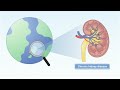
Li Y, Waheed YA, Sun D
International Journal of Nanomedicine 2025, 20:11267-11294
Published Date: 13 September 2025