Back to Journals » Nanotechnology, Science and Applications » Volume 19
Nanostructured Lipid Carrier-Gels for Wound Healing: A Narrative Review of Formulation Strategies, Mechanisms, and Translational Potential
Authors Alquraisy A, Ramadhani K, Mohammed AFA, Wilar G, Osman-W, Elamin KM ![]() , Wathoni N
, Wathoni N ![]()
Received 28 November 2025
Accepted for publication 23 January 2026
Published 18 February 2026 Volume 2026:19 585159
DOI https://doi.org/10.2147/NSA.S585159
Checked for plagiarism Yes
Review by Single anonymous peer review
Peer reviewer comments 4
Editor who approved publication: Professor Kattesh Katti
Ayatulloh Alquraisy,1 Khoirunnisa Ramadhani,1 Ahmed Fouad Abdelwahab Mohammed,2,3 Gofarana Wilar,4 Wadah Osman,5 Khaled M Elamin,6 Nasrul Wathoni1
1Department of Pharmaceutics and Pharmaceutical Technology, Faculty of Pharmacy, University of Padjadjaran, Sumedang, 45363, Indonesia; 2Department of Pharmaceutics, Faculty of Pharmacy, Minia University, Minia, 61519, Egypt; 3Department of Pharmaceutics, Faculty of Pharmacy, Minia National University, Minia, 61768, Egypt; 4Department of Pharmacology and Clinical Pharmacy, Faculty of Pharmacy, University of Padjadjaran, Sumedang, 45363, Indonesia; 5Department of Pharmacognosy, Faculty of Pharmacy, Prince Sattam Bin Abdulaziz University, Al-kharj, Saudi Arabia; 6Graduated School of Pharmaceutical Science, Kumamoto University, Kumamoto, 862-0973, Japan
Correspondence: Nasrul Wathoni, Email [email protected]
Abstract: Wound healing is a complex process involving hemostasis, inflammation, proliferation, and remodeling of tissues or cells. In chronic conditions, such as diabetic wounds, this process is often disrupted. Nanostructured lipid carriers (NLCs) are advantageous topical drug delivery systems because of their ability to improve stability, bioavailability, and controlled drug release. The incorporation of NLCs into a gel matrix (NLC-gel) enhances the formulation with bioadhesive properties, skin hydration, and drug retention in the wound area. This accelerates healing and reduces the risk of infections. While these findings highlight the potential of NLC–gel systems to improve local drug bioavailability and promote tissue regeneration, most available evidence is derived from in vitro and animal studies, and clinical data remain limited. This review critically summarizes recent advances in NLC–gel formulations for wound healing, with particular emphasis on the relationship between formulation strategies and biological mechanisms, including modulation of inflammation, angiogenesis, fibroblast and keratinocyte proliferation, collagen deposition, and antimicrobial and antioxidant activities. Additionally, translational considerations such as long-term safety, formulation scalability, and clinical prospects are discussed. Recent in vivo and in vitro studies have shown that NLC gels containing active ingredients, such as simvastatin, curcumin, quercetin, moxifloxacin, or other therapeutic proteins, can accelerate wound healing, particularly in wounds caused by metabolic disorders. These results suggest that NLC gels have great potential as therapeutic platforms for wound care. However, further research is required to optimize its formulation and clinical translation.
Keywords: nanostructured lipid carriers, gel, hydrogel, wound healing
Introduction
Wound healing is a tightly regulated biological process involving hemostasis, inflammation, proliferation, and tissue remodeling to replace damaged components. Anti-inflammatory agents are used to reduce complications caused by an excessive immune response at the wound site.1 This process involves the migration and proliferation of blood cells, production of extracellular matrix, endothelial cells, cytokines, release of growth factors, angiogenesis, and fibroblasts.2 Pharmacological agents can aid in accelerating the wound-healing process.3,4 Acute wounds generally progress through the healing phases in a timely and coordinated manner, whereas chronic wounds are characterized by delayed or arrested healing, often associated with persistent inflammation, impaired angiogenesis, and metabolic disorders such as diabetes, making them the primary target for advanced drug delivery approaches, including nanocarrier-based systems.5,6 Conventional topical wound treatments often face limitations such as poor drug penetration and stability, rapid uncontrolled release, low wound retention, microbial colonization, oxidative stress, and inadequate vascularization. Topical drug delivery systems enable localized and sustained drug release at wound sites, reducing systemic effects. However, conventional hydrophilic bases often fail to maintain moisture and control drug release, resulting in frequent reapplication, suboptimal local concentrations, and inconsistent healing outcomes.7–9 Conventional gels provide hydration and ease of use but lack controlled drug release, offer limited protection against degradation, and are ineffective for delivering lipophilic drugs, reducing their therapeutic potential in complex wounds.10,11 Beyond hydrogel-based matrices, decellularized extracellular matrix (dECM) scaffolds have emerged as bioactive platforms for wound repair and tissue regeneration, owing to their preserved structural, biochemical, and signaling components that actively regulate cell migration, angiogenesis, and tissue remodeling.12 Recent advances in dECM processing and functionalization further highlight its role as a benchmark scaffold in tissue engineering, providing a useful reference framework for evaluating synthetic and hybrid delivery systems, including NLC–gel formulations. Therefore, nanocarrier-based topical systems, especially nanostructured lipid carriers (NLCs), offer improved bioavailability, drug protection, and tissue repair. Their mixed solid–liquid lipid matrix provides higher drug loading capacity and lower drug expulsion compared to solid lipid nanoparticles (SLNs).13 NLC increase the stability and effectiveness of drugs by protecting them, increasing drug loading capacity, controlling drug release, improving drug release kinetics, and facilitating better absorption.11,14 When incorporated into gel formulations, NLCs further enhance skin hydration and adhesion, protect the drug within the lipid matrix, and enable direct delivery to the wound site. NLC-based gels maintain local drug concentrations for a longer period, reduce systemic absorption, and provide anti-inflammatory, antioxidant, and pro-healing effects, making them a promising strategy for improving wound healing outcomes.9,15 Several review articles have previously discussed lipid-based nanocarriers, including solid lipid nanoparticles (SLNs) and nanostructured lipid carriers, for wound healing applications. However, most of these reviews either broadly cover multiple nanocarrier systems or focus predominantly on formulation aspects without critically linking formulation parameters to biological mechanisms and therapeutic outcomes. In addition, the specific advantages and challenges of NLC-based gel systems, particularly regarding wound retention, local bioavailability, mechanistic pathways, and translational potential, remain insufficiently explored. Therefore, the present review aims to fill this gap by providing a focused and mechanistically oriented overview of NLC–gel systems for wound healing, integrating formulation strategies, physicochemical and biological characterization, mechanistic insights, and emerging therapeutic directions.
Nanostructured Lipid Carriers (NLCs): Fundamentals and Applications
Composition and Key Components of NLCs
Nanostructured Lipid Carriers (NLCs) are advanced lipid-based nanocarriers composed of a mixture of solid and liquid lipids stabilized by suitable surfactants. The combination of solid and liquid lipids forms an imperfect crystalline matrix that enhances the drug loading capacity and prevents drug expulsion during storage, which is a limitation of first-generation solid lipid nanoparticles (SLNs).16,17 Solid lipid components, such as glyceryl monostearate, stearic acid, and beeswax, provide structural integrity and modulate the release rate of the encapsulated drug.18,19 Liquid lipids, such as oleic acid, medium-chain triglycerides, and natural oils, introduce structural imperfections into the solid matrix, improving drug accommodation and controlling polymorphic transitions.20 This can be combined with liquid lipids or oils (eg, argan oil, oleic acid, medium-chain triglycerides) to create the structural “imperfections” necessary for higher encapsulation efficiency of lipophilic active ingredients.21 The selection of surfactants is crucial for the formation of NLCs to obtain a skin application size range (150–300 nm), colloidal stability, and biocompatibility. Surfactants and co-surfactants, such as Tween 80, Poloxamer 188, and lecithin, stabilize the dispersion and influence the droplet size and zeta potential.22 In addition, there are polysorbates for topical and dermal applications, sucrose esters (sucrose monostearate (SS) and sucrose monopalmitate (SM)) as biodegradable non-ionic surfactants, so they are better tolerated and less irritating than polyethoxylated surfactants, α-Tocopheryl Acetate (αTA) to chemically stabilize lipids, pentylene glycol (PG) as a preservative to ensure antimicrobial stability of the formulation, with good skin tolerability and suitability of NLC formulations.23,24 The proper selection of these components determines the physicochemical stability, entrapment efficiency, and release kinetics of NLC systems.25 Analysis of the formulations summarized in Table 1 reveals a consistent preference for mixed solid–liquid lipid matrices, typically combining lipids such as glyceryl behenate, precirol ATO 5, stearic acid, or glycerol monostearate with oils including medium-chain triglycerides, oleic acid, or Miglyol derivatives. This strategy is applied across a wide range of wound-healing actives, indicating its versatility for topical delivery. Non-ionic surfactants, particularly Tween 80, Poloxamers, and Kolliphor derivatives, dominate the formulations, reflecting their favorable skin compatibility, although surfactant selection and ratios vary considerably among studies. Carbopol-based hydrogels emerge as the most frequently used gel matrix, followed by thermoresponsive poloxamer and chitosan-based systems, highlighting the importance of tunable rheology and bioadhesion for wound applications. Preparation methods are predominantly based on hot homogenization combined with sonication, with some studies incorporating experimental design approaches to optimize formulation variables. Overall, while recurring formulation trends are evident, the heterogeneity in formulation composition and processing underscores the need for more standardized, design-driven strategies to improve comparability and translational relevance.
 |
Table 1 Formulation Components And Preparation Methods Of NLC-Gel Systems For Wound Healing Applications |
Mechanisms of Enhanced Drug Loading, Controlled Release, and Skin Permeation
The inclusion of solid and liquid lipids allows NLCs to encapsulate a wide range of hydrophilic and lipophilic drugs with an enhanced entrapment efficiency compared to solid lipid nanoparticles.39 NLCs are typically composed of a mixture of solid and liquid lipids, resulting in a less ordered (amorphous) core compared to fully crystalline solid lipid nanoparticles (SLNs). This disordered matrix provides more space (imperfections) to accommodate drug molecules. Amorphous regions in the lipid core reduce drug crystallization, allowing for a higher loading capacity and prolonged release.40,42 Controlled release is achieved through lipid diffusion and degradation, which can be modulated by the lipid composition and surfactant concentration. Controlled and sustained release from NLCs arises from the slow diffusion of drug molecules through the lipid matrix and the gradual reorganization/crystallization of lipids, which has been shown to prolong local drug availability in wound models.15,16 Mechanistically, the nanometric size (typically < 200 nm) and lipid composition of NLCs may enhance skin permeation and surface area by increasing drug partitioning into skin lipids and temporarily disrupting the stratum corneum lipid domain processes, as demonstrated in ex vivo permeation and in vivo wound studies, as well as promoting passive diffusion of the drug across the skin barrier.34,43 The nanoscale size of NLCs increases their surface area and allows direct contact with the skin surface, facilitating a higher concentration gradient and enhancing the drug flow into the skin.44 The lipid composition of NLCs not only protects labile bioactives from environmental degradation (oxidation and enzymatic hydrolysis) but also imparts an occlusive effect on lipid nanoparticles, which improves skin hydration and permeability by reducing transepidermal water loss and helps maintain a moist wound microenvironment that supports re-epithelialization. This occlusion slows drug release from the skin surface, effectively creating a reservoir. Increased hydration can disrupt the dense lipid structure of the stratum corneum, thereby increasing its permeability.11,45 Together, these mechanisms ensure the sustained and localized delivery of therapeutic molecules directly to the wound site.21,30
Pharmaceutical and Herbal Compounds for Wound Healing: Rationale for Incorporation into NLC–Gel Systems
Various bioactive compounds have been successfully incorporated into NLCs for wound-healing applications. Synthetic drugs, such as doxycycline hydrate,32 simvastatin,34 phenytoin,30 and lidocaine,33 have demonstrated enhanced dermal retention and controlled release from NLC formulations. Natural compounds and herbal active ingredients, including curcumin, resveratrol,19 aloin, astaxanthin,7 zerumbone,15,22 and rosemary essential oil,39 have demonstrated potent antioxidant and anti-inflammatory properties when delivered via NLCs. Curcumin-loaded NLCs accelerated full-thickness wound closure and reduced oxidative/inflammatory markers in animal models, whereas simvastatin-loaded NLC gels improved ulcer healing and histological regeneration in vivo.34,43 In addition, the encapsulation of propolis37 and St. John’s Wort extracts26 in lipid carriers enhanced antimicrobial activity and promoted faster epithelialization than free extracts. Many of these compounds exhibit high lipophilicity, poor aqueous solubility, and susceptibility to oxidative or enzymatic degradation, which limit their effectiveness in conventional hydrophilic gel formulations. Encapsulation within the imperfect lipid matrix of NLCs enhances drug solubilization, protects labile compounds from degradation, and enables sustained release at the wound site. The gel matrix further complements NLCs by improving wound adhesion, hydration, and local drug retention, thereby creating a synergistic delivery platform that enhances the therapeutic performance of both pharmaceutical and herbal bioactives in complex wound environments. Finally, recent primary research has demonstrated that formulation variables (lipid type, surfactant concentration, and production method) and processing conditions (homogenization/sonication parameters and cooling profile) critically determine NLC physicochemical attributes and their subsequent wound-healing performance, which underscores the importance of systematic optimization in translational development and the versatility of NLCs in delivering pharmaceutical and phytochemical agents for comprehensive wound management.46
NLC–Gel Hybrid Systems for Topical Wound Healing
Role of Gel Matrices in NLC-Gel Hybrid Systems
Gel matrices are widely used in topical wound therapy due to their ability to form hydrophilic three-dimensional networks that maintain a moist wound environment. As semisolid systems, gels provide appropriate viscosity and spreadability.29,46 Common polymers such as carbopol, poloxamer, sodium alginate, and chitosan17,25 form cross-linked networks capable of retaining large amounts of water and therapeutic agents, thereby sustaining the microenvironment required for effective wound healing.31,47 Carbopol-based hydrogels are particularly valued for their transparency and spreadability, whereas poloxamers exhibit thermoresponsive sol–gel transition at skin temperature, facilitating in situ application and improved retention.48 In addition, natural polymer-based gels such as chitosan and alginate contribute intrinsic biocompatibility and biological activity, including antimicrobial and hemostatic properties, which further support wound repair.49 From a formulation perspective, gel matrices also offer practical advantages as topical dosage forms. Their semisolid consistency improves handling, ease of application, and dose reproducibility compared with liquid dispersions, while reducing discomfort during application and removal.19 The bioadhesive nature of gels prolongs residence time at the application site and minimizes dosing frequency, contributing to improved patient compliance.38,40 Hydrogels maintain sustained hydration at the wound surface, which favors fibroblast migration and granulation tissue formation,21 and often exhibit shear-thinning behavior that facilitates spreading while allowing the formulation to remain in place after application.50 Moreover, the hydrophilic polymer network protects incorporated actives from environmental and thermal degradation, enabling the inclusion of thermolabile compounds and improving formulation stability during storage.22,25 Gel matrices also reduce physical instability such as aggregation and sedimentation, resulting in more stable and user-acceptable topical systems.46 Beyond these vehicle- and dosage-form-related functions, the integration of nanostructured lipid carriers within gel matrices generates synergistic effects that enhance therapeutic performance beyond that achievable by either component alone.18,28 The gel network immobilizes NLCs, improving mechanical stability and enabling more controlled drug release at the wound site.51 This synergistic interaction supports sustained local bioavailability and prolonged wound contact, translating into enhanced wound healing outcomes compared with gel-based or NLC-based systems alone. In vivo studies consistently report accelerated wound closure and improved tissue regeneration with NLC–gel formulations, highlighting the therapeutic advantage of this hybrid approach.33,52 Importantly, the extent of synergy depends on the choice of gel polymer, as polymer composition influences mechanical strength, release kinetics, and permeation behavior; therefore, gel selection should be tailored to the intended wound environment and therapeutic objective.53
Formulation Strategies
Analysis of NLC-Gel and Other Topical Nanocarriers
Nanostructured lipid carriers (NLCs) are versatile topical systems for wound healing, offering enhanced drug stability, controlled release, higher drug loading, and reduced drug expulsion compared to solid lipid nanoparticles (SLNs), while also improving hydration and adhesion to support sustained therapeutic levels and faster tissue repair. In addition to lipid carriers like NLCs, other topical nanocarriers such as polymeric nanoparticles (PNPs), nanoemulsions, and ethosomes have been extensively investigated for wound healing applications. A semi-quantitative comparison of topical nanocarrier systems based on particle size and entrapment efficiency (EE) revealed formulation- and manufacturing-method-dependent differences relevant to wound healing performance. Nanostructured lipid carriers (NLCs) typically exhibit particle sizes in the range of approximately 80–300 nm, with high entrapment efficiencies generally reported between 70% and 95%, due to the imperfect solid-liquid lipid matrix, which allows for higher drug loading and minimizes unwanted release during storage.54 Solid lipid nanoparticles (SLNs) typically exhibit particle sizes in the range of approximately 100–1000 nm, with more variable and often lower %EE values, typically in the range of 50–85%, due to lipid crystallization and polymorphic transitions that can promote drug release over storage.54,55 Polymer nanoparticles (PNPs), such as chitosan- or PLGA-based systems, typically range in size from approximately 100–400 nm and exhibit moderate to high entrapment efficiencies, generally reported between 60–90%, depending on the polymer composition, molecular weight, and drug-polymer interactions.56
Nanoemulsions generally have droplet sizes of approximately 100–200 nm and exhibit good solubilization capacity for lipophilic drugs, with reported %EE values typically ranging from 60–85%. However, the absence of a solid matrix results in faster drug diffusion and less controlled release compared to lipid nanoparticle systems, although their ability to enhance skin permeation has been well documented.57 Ethosomes, which consist of ethanol-rich phospholipid vesicles, typically have particle sizes between approximately 120–400 nm with %EE values often exceeding 70–90%, and are highly effective in enhancing dermal and transdermal penetration, although formulation durability and potential ethanol-related irritation remain issues to consider.54,58 Overall, this semi-quantitative comparison demonstrates that while SLNs, PNPs, nanoemulsions, and ethosomes each offer specific advantages, NLC-based systems provide a more balanced combination of nanoscale size, consistently high capture efficiency, and controlled release behavior, supporting their growing relevance in advanced topical wound healing strategies.
Polymeric nanoparticles, composed of biodegradable polymers such as PLGA, chitosan, or collagen, can encapsulate therapeutic agents to protect them from premature degradation and provide controlled release, often resulting in improved drug stability and retention at the wound site with relatively high loading capacities; however, their formulation typically involves complex synthesis steps and purification that can increase production costs and manufacturing complexity, which has hindered clinical translation despite strong preclinical interest.59,60 Nanoemulsions are oil–water dispersions stabilized by surfactants that enhance skin permeation by altering barrier properties and can be simpler and cheaper to produce than polymeric systems; they also offer moderate drug loading and kinetic stability, though they may be prone to physical instability (eg, droplet coalescence) under certain conditions and may require additional gelling agents to improve retention at wound sites.61 Ethosomes are soft phospholipid vesicles containing high concentrations of ethanol, increasing lipid fluidity and facilitating deeper penetration of both hydrophilic and lipophilic drugs through the stratum corneum, often outperforming conventional liposomes in skin delivery; while ethosomes can achieve high entrapment efficiency and enhanced penetration, the presence of ethanol can pose formulation challenges such as potential irritation and requires careful optimization, and their manufacturing is generally more complex than that of simple nanoemulsions.45 Collectively, these platforms demonstrate varied advantages and limitations: polymeric nanoparticles excel in controlled release and stability but at higher cost and complexity, nanoemulsions offer ease of production and good penetration enhancement with moderate stability, and ethosomes provide superior permeation at the expense of formulation challenges and potential irritation. All of which should be weighed when selecting a nanocarrier for topical wound healing.
Lipid and Surfactant Selection Strategy
The selection of appropriate lipids and surfactants is crucial for developing stable and effective NLC formulations and creating an imperfect crystalline matrix that maximizes encapsulation of the intended active molecule.17 The goal is to select a solid and liquid lipid that can form a stable NLC matrix and effectively dissolve the active pharmaceutical ingredient (API).62 Screening is typically performed by evaluating the solubility of the active compound in various solid and liquid lipids, such as glyceryl monostearate, stearic acid, beeswax, oleic acid, and caprylic triglyceride, by measuring drug solubility/partitioning in candidate lipids, thermal behavior (DSC), and solubility of solid/liquid lipid mixtures because drug solubility in the lipid matrix significantly determines entrapment and release efficiency.18,19 Experimental studies periodically performed solubility testing (drug in lipid), DSC/pXRD to check crystallinity, and short-term stability screening to select the optimal lipid mixture.63,64 Surfactants and co-surfactants are essential for stabilizing NLC dispersions and lowering interfacial tension and HLB. Various surfactants/co-surfactants were tested for their ability to form a stable pre-emulsion with the selected lipid mixture and aqueous phase.65 Surfactants such as Poloxamer 188, Tween 80, and lecithin were selected based on their ability to maintain colloidal stability during homogenization or ultrasonication and to achieve a stable nanoparticulate dispersion with the desired particle size and zeta potential for topical application, emulsification ability, skin toxicity/compatibility, and the hydrophile-lipophile balance (HLB) required to form stable NLC nanoemulsions/dispersions.47,66 The combination of solid and liquid lipids, along with the choice of surfactant, determines the particle size, zeta potential, and encapsulation efficiency. Compatibility studies are often performed to ensure chemical stability and to prevent phase separation or drug leakage over time.20 The visual representation of the NLC formulation loaded into the gel is shown in Figure 1.
 |
Figure 1 NLC consists of a solid–liquid lipid matrix stabilized by surfactants, enabling high drug loading and controlled drug release.67 Embedded within a hydrogel network, the NLC-gel provides enhanced skin penetration, prolonged retention, and improved wound-healing performance.47 |
Application of Experimental Design in NLC Optimization
NLC formulation parameters are generally optimized using statistical tools such as Design of Experiment (DoE) or Response Surface Methodology (RSM), which allow for the systematic evaluation of variables such as lipid ratio, surfactant concentration, and homogenization rate.25 The widespread use of DoE in early research involved factorial designs, Box-Behnken designs (BBD), Central Composite Designs (CCD), and D-optimal designs to study factors such as lipid percentage (solid:liquid ratio), surfactant concentration, sonication/homogenization time, and temperature.68,69 This approach efficiently identifies the critical formulation and process parameters, minimizes the number of experiments while identifying the optimal formulation with the desired particle size, polydispersity index (PDI), and entrapment efficiency, and helps achieve the desired product quality attributes at a lower cost and time than traditional methods.70 Some experimental groups combine elements of QbD (defining CQAs, risk assessment, and DoE) to allow optimization to be transferred to scale-up and to define design space boundaries for process robustness.71 For example, an optimized NLC-gel formulation of doxycycline and aloin achieved a uniform size distribution and high drug loading, thereby accelerating the healing of diabetic wounds. Similarly, a curcumin–resveratrol combination NLC gel developed using RSM demonstrated enhanced antioxidant activity and tissue regeneration in skin wounds. Thus, statistical optimization ensures the production of reproducible and scalable, high-performance NLC systems. Processing parameters, such as high-pressure homogenization or probe sonication, temperature during emulsification, and cooling profile during lipid solidification, strongly influence the particle size distribution, polydispersity, and entrapment efficiency, and must be systematically optimized.19,22
Incorporation of Active Pharmaceutical Ingredients into NLC Systems
The incorporation of actives into a nanostructured lipid carrier (NLC) gel involves a two-step process: first, loading the active agent into the NLC particles and second, dispersing these loaded nanoparticles into a gel base.72 Various preparation methods have been developed for nanostructured lipid carriers (NLCs), and the choice of technique strongly influences particle characteristics, drug loading, and suitability for topical gel formulations. High-pressure homogenization, in both hot and cold variants, is the most widely used method due to its scalability, solvent-free processing, and ability to produce particles with narrow size distributions; however, exposure to elevated temperatures in the hot process may compromise thermolabile active ingredients.73 Melt emulsification represents a simple and reproducible approach that avoids organic solvents, although it may result in broader particle size distributions and limited control over lipid polymorphism.73 Solvent-based methods, such as emulsification–evaporation or diffusion, enable efficient incorporation of poorly water-soluble drugs at relatively low processing temperatures but require careful removal of residual solvents to ensure safety and regulatory compliance.73 Microemulsion-based techniques offer excellent control over particle size through the formation of thermodynamically stable precursors, yet they often rely on high concentrations of surfactants and co-surfactants, which may limit their applicability in topical formulations.74 Phase inversion methods, including phase inversion temperature approaches, allow NLC formation with relatively low energy input and are particularly attractive for heat-sensitive compounds; nevertheless, these methods typically exhibit narrow processing windows and strong dependence on formulation composition.74 Collectively, these preparation strategies, as summarized in recent comprehensive reviews, highlight the need for rational method selection based on the physicochemical properties of the active ingredient and the intended performance of NLC-based gel systems.73,74
Herbal active ingredients such as propolis, rosemary essential oil, and St. John’s Wort extract, have been successfully integrated into NLCs to maintain biological activity and enhance penetration into deeper skin layers.37,39 For example, zerumbone-loaded NLCs and astaxanthin-loaded chitosan-coated NLCs exhibited enhanced antioxidant and anti-inflammatory effects in a diabetic wound model.7,15 Appropriate incorporation techniques are crucial for achieving homogeneous drug distribution, high entrapment efficiency, and stability during storage.32 The polarity and solubility of an active compound determine the optimal lipid composition and preparation method. NLCs protect the incorporated active ingredients from chemical degradation (eg, hydrolysis and oxidation), thereby extending the product shelf life. The NLC dispersion is physically compatible with gel excipients (polymers, surfactants, etc.) to prevent the aggregation of nanoparticles or other instabilities.75 While formulation strategies define the rational design and compositional framework of NLC–gel systems, they do not inherently guarantee optimal biological performance. The translation of lipid selection, surfactant optimization, and processing conditions into therapeutic efficacy requires systematic physicochemical and mechanical characterization. Therefore, characterization parameters serve as a critical bridge between formulation design and biological function, providing measurable indicators of stability, skin interaction, drug release behavior, and wound-healing potential. In this context, parameters such as particle size, surface charge, entrapment efficiency, morphology, and gel rheology are not merely descriptive attributes, but functional determinants that govern the in vivo performance of NLC–gel systems.
Characterization Parameters
Particle Size, Polydispersity Index, and Zeta Potential
Particle size, polydispersity index (PDI), and zeta potential are the primary characterization metrics because they correlate with colloidal stability and skin interaction.46,76 The particle size and Polydispersity Index are important indicators of NLC uniformity and stability, which are usually measured by dynamic light scattering (DLS), and are critical because smaller, uniform nanoparticles favor good skin penetration, stability, and controlled release.17 Optimal wound-healing formulations typically exhibit particle sizes below 200 nm with narrow PDI values (<0.3), thus ensuring better penetration and minimal aggregation.52 For example, simvastatin-loaded NLCs used for wound healing have an average diameter of ~100 nm.34 The zeta potential reflects the surface charge and electrostatic stability of the nanoparticles; values greater than ±30 mV are generally considered stable against aggregation.25 For example, zerumbone-loaded NLC gels exhibit a zeta potential of approximately −35 mV, which contributes to their excellent colloidal stability and sustained release.15,22 In NLC studies, the influence of process parameters (lipid composition, homogenization, etc.) on particle size and stability was characterized by dynamic light scattering (DLS), which showed that particle sizes of ~60–80 nm and zeta potential reaching ~−30 mV indicated good colloidal stability.77 Another formulation (NLC-containing albendazole) reported optimized particles with a size of ~176.5 nm, a PDI of 0.41, and a zeta potential of −22.7 mV, indicating a relatively narrow size distribution and stable charge.78 The physicochemical data summarized in Table 2 indicate that most NLC–gel formulations for wound healing exhibit particle sizes predominantly below 200 nm with relatively narrow polydispersity indices (PDI < 0.3), reflecting generally uniform nanoscale dispersions suitable for topical application. Entrapment efficiency values are consistently high across studies, frequently exceeding 80% and in several cases approaching or surpassing 90%, particularly for lipophilic and phytochemical actives. These findings support the suitability of NLC-based systems for efficient drug incorporation within semisolid topical matrices. Zeta potential values span a broad range from moderately negative to strongly positive, indicating diverse stabilization strategies depending on lipid composition and surfactant selection. Notably, both negatively and positively charged systems demonstrate high entrapment efficiency and acceptable particle size distributions, suggesting that surface charge alone does not dictate encapsulation performance or dispersion quality. Morphological analyses, where reported, consistently describe spherical and smooth nanoparticles, further supporting the physical integrity of NLCs within gel systems. However, the lack of standardized reporting particularly for long-term stability and complete surface charge characterization limits direct comparison across studies and highlights the need for harmonized physicochemical evaluation in future formulation design.
 |
Table 2 Physicochemical Characterization Parameters Of NLC-Gel Formulations For Wound-Healing Applications |
Entrapment Efficiency and Drug Loading
Entrapment efficiency (EE%) and drug loading (DL%) were measured to quantify the extent of activity associated with the lipid matrix versus free in the aqueous phase, and these values directly influenced the release kinetics and in vivo efficacy.80 The entrapment efficiency (EE) and drug loading (DL) indicate the proportion of active compounds successfully encapsulated in the lipid matrix.30 High EE values (>80%) are desirable for sustained release and reduced wastage of bioactive ingredients.27 The lipid composition, surfactant concentration, and preparation method significantly influenced these parameters. In NLCs made with different liquid lipid types (eg, black seed oil and coconut oil), researchers compared entrapment efficiency among formulations, measured EE, and drug loading, and found that the choice of lipid significantly influenced EE.81 In the albendazole-NLC system (RSC study), the entrapment efficiency reached ~89.85%, and the drug loading capacity was ~3.19%, indicating that a high proportion of the drug can be stably incorporated.78 Studies involving simvastatin and doxycycline have reported high EE and DL values due to the partial crystallinity of NLCs, which provides sufficient space for drug incorporation.32,52 Maintaining high entrapment while preventing burst release is crucial for achieving prolonged therapeutic effects in the wound environment.
Morphological Characterization (SEM/TEM)
Morphological characterization using Scanning Electron Microscopy (SEM) or Transmission Electron Microscopy (TEM) confirmed the shape, surface texture, surface characteristics, and structural integrity of NLCs, which can help explain the release phenomena and skin deposition observed in ex vivo and in vivo studies, thereby ensuring a stable and effective drug delivery system.46,82 Most wound-healing NLCs exhibited a spherical or oval morphology with smooth surfaces, indicating successful lipid stabilization and minimal aggregation. For instance, melatonin-loaded microspheres29 and phenytoin-loaded30 NLCs exhibited uniform spherical shapes, which correlated with their controlled drug release profiles and improved bioadhesion on to wound bed. TEM images often reveal a dense core–shell structure, confirming the encapsulation of bioactive compounds within the lipid matrix.39 In the clobetasol propionate NLC gels, SEM was used to observe the morphology, which predominantly exhibited spherical to slightly oval shapes, indicating the formation of uniform lipid droplets within the nanostructured matrix. The particles appeared to have a smooth surface, indicating the proper encapsulation of clobetasol propionate within the lipid mixture without surface crystallization or drug efflux.83 In the curcumin-NLC formulation for skin regeneration, SEM showed an irregular spherical shape, and TEM confirmed the nanoparticle structure, supporting a uniform morphology.43
Gel Properties and Rheological Behavior
The physicochemical evaluation of NLC gels ensures compatibility with skin physiology and user acceptability.38 The ideal pH range (5.5–7.0) prevents irritation while maintaining the stability of the drug. Viscosity influences the retention time and release rate of the drugs. Liquid NLCs are formulated into semi-solid forms (gel formulations) because of their low viscosity and poor adhesiveness upon topical application. An appropriate concentration of the gelling agent was selected to provide optimal organoleptic properties and viscosity was selected.84 Formulations using carbopol or chitosan exhibit shear-thinning behavior, which is suitable for topical applications.31 Spreadability and extrudability determine the ease of application and the uniform distribution over the wound site.25 Texture profile analysis provided insights into the firmness, cohesiveness, and adhesiveness, which directly affect the bioadhesive performance of NLC gels. Overall, these parameters ensured optimal consistency, aesthetic appeal, and therapeutic efficiency of the final formulation.18,30 In a study of ibuprofen-containing NLC gels, Texture Profile Analysis (TPA) was used to measure hardness, adhesion, cohesiveness, and firmness, which are highly relevant to the texture profile. A reverse extrusion test was also performed to assess extrudability, with spreadability measured using a more precise instrument-based texture analyzer that correlated with firmness and consistency.85 In a study of NLC-mediated transdermal aceclofenac hydrogels, the texture properties were characterized using a texture analyzer, and firmness (gel strength), spreadability (shearing effort), and adhesion (adhesion effort) were measured to remove the texture probe.86 In a recent study on amphotericin B-containing NLC gels, pH, extrusion ability, and spreadability were measured. A gel with a pH of 6.3, which is relatively skin-friendly, extrudability of 9.5 ± 0.12 g/cm2, and spreadability of ~6.4 cm2 was produced.87
Mechanisms of Enhanced Wound Healing
Sustained Drug Release and Prolonged Local Bioavailability
Nanostructured lipid carriers (NLCs), bigels, and hybrid hydrogels enhance wound healing by providing sustained and controlled release of active compounds. This mechanism reduces sudden release, ensuring that therapeutic molecules remain at the wound site for a longer period, reducing the need for repeated applications while maintaining effective drug concentrations, including drugs such as simvastatin,34 phenytoin,30 melatonin,29 astaxanthin,7 zerumbone,22 and St. John’s Wort extract.26 This controlled release accelerated re-epithelialization or tissue repair, enhanced angiogenesis, and increased collagen synthesis/deposition in both acute and chronic wounds. In preclinical biodistribution studies, topically administered NLCs remained localized in the skin for up to 24 h with minimal systemic absorption, preventing nanoparticle penetration, thus supporting a prolonged and safe local effect after topical application to wounds.88 In another study, NLC encapsulated hydrophobic and hydrophilic active ingredients in a solid-liquid lipid matrix containing epidermal growth factor and curcumin. The results showed that NLC can slow diffusion, protect the payload from rapid clearance or degradation, accelerate wound closure, and enhance the antioxidant enzyme activity. Thus, it has the potential to accelerate the healing of chronic wounds.89 When integrated into gels, such as carbopol-based hydrogels or collagen/chitosan scaffolds, the lipid + polymer gel matrix provides a depot effect that releases the drug over hours to days, maintaining therapeutic concentrations in the wound bed, reducing dosing frequency, and increasing tissue exposure for repair.90 It should be noted that the majority of biological effects observed in NLC–gel formulations arise from the pharmacological activity of the incorporated agents, while the primary contribution of the NLC–gel carrier lies in enhancing drug stability, local bioavailability, and sustained exposure at the wound site, thereby potentiating these effects rather than acting as a therapeutic entity on its own.
Enhanced Skin Penetration and Dermal Drug Deposition
Many drugs experience limited skin permeation because of the barrier properties of the stratum corneum. NLCs enhance drug permeation by mimicking the lipid architecture of the skin through the combination of solid and liquid lipids, thereby increasing fluidity and dermal absorption. Enhanced skin penetration and dermal deposition in NLCs and hybrid hydrogels as visualized in Figures 2 and 3 have been shown to be effective demonstrated for difficult-to-administer combination drugs such as curcumin,46 quercetin-resveratrol,19 astaxanthin,7 mupirocin,35 doxycycline,32 and lidocaine.33 Enhanced systems allow drugs to pass through the stratum corneum or reach deeper tissue layers more effectively by utilizing the occlusive effect of small particle which enhance hydration and transfollicular entry. This allows small molecules and phytochemicals to reach deeper dermal layers, where inflammation, bacterial infection, and impaired angiogenesis commonly occur. This could result in improved therapeutic strategies for wound healing. Nanometric lipid particles (NLCs) interact with stratum corneum lipids, disrupting the barrier, and act as penetration enhancers by enhancing drug partitioning and retention within the epidermis/dermis. Small particle size and lipid composition (solid + liquid lipids, surfactants) support closer contact and better dermal absorption than conventional formulations. In NLC gel studies using lidocaine, higher dermal deposition and better local effects were demonstrated when NLC was administered owing to increased skin permeation and higher local drug levels.91 In NLC studies using idebenone, significant improvements in stability and skin permeation were achieved, thus potentially becoming an alternative wound treatment.92
 |
Figure 2 Formation of NLCs, their incorporation into hydrogels to create NLC-gels, and the resulting enhancement in wound healing through improved penetration, sustained drug release, and accelerated tissue repair.51 The upward arrows indicate enhancement of key wound healing processes, including improved cellular infiltration, angiogenesis, and tissue regeneration at the wound site. |
 |
Figure 3 Comparative skin penetration profile of free drug, nanostructured lipid carriers (NLC), and NLC-gel.93 Free drugs show minimal and superficial penetration due to rapid clearance and low affinity for the stratum corneum. NLC enables deeper dermal penetration through lipid–lipid compatibility and nanoparticle-mediated transport.94 NLC-gel provides the most optimal penetration and retention due to its combination of nanoscale penetration enhancement and hydrogel-mediated depot effect, resulting in prolonged drug availability at the wound site.51 |
Anti-Inflammatory, Antioxidant, and Antimicrobial Mechanisms
Many nanoformulations possess anti-inflammatory, antioxidant, and antimicrobial activities, regardless of the drug itself (quercetin, resveratrol,19 and mupirocin),35 natural active substances (rosemary essential oil,39 thymol,40 propolis,37 and mentha pulegium oil38), or lipid excipients, which contribute to wound infection control by suppressing colonization, inhibiting microbial growth, and reducing oxidative stress. Thus, these mechanisms can synergistically accelerate epithelialization, reduce cytokine levels, collagen deposition, and granulation tissue formation, and accelerate the healing of infected or chronic wounds. Furthermore, the combination of NLCs with biologics (eg, growth factors) and polymeric scaffolds potentiated neovascularization and organized matrix formation in full-thickness models.95 NLC gel delivery with anti-inflammatory active ingredients (zerumbone,15 simvastatin,34 and pioglitazone) resulted in a greater reduction in inflammatory markers and faster histological wound repair in animal wound models, consistent with better local availability and sustained exposure. NLC, which simultaneously deliver antioxidants (curcumin and growth factors), increase the activity of antioxidant enzymes (SOD, catalase, GPx), which contribute to accelerated healing and improved tissue remodeling.89 The controlled release of NLC helps to maintain antioxidant levels in the wound. Topical antimicrobials incorporated into NLC demonstrated improved wound retention and sustained antibacterial action against wound pathogens, resulting in better infection control. The composition of the NLC (certain lipids/surfactants) may also contribute to membrane interactions that enhance antimicrobial efficacy.96
Evaluation Models
In vitro Evaluation of Cytocompatibility and Wound-Healing Activity
In vitro studies are essential for validating the safety and wound-healing potential of this formulation. These tests include cell viability, scratch assay (cell migration), DAPI staining, and cell cycle analysis (assessing cell proliferation), which are commonly used to evaluate the cytocompatibility of NLC formulations containing genistein-dexamethasone-moxifloxacin,27 curcumin-resveratrol,79 simvastatin,34 astaxanthin,7 and recombinant thrombomodulin.17 This ensured their regenerative potential before in vivo testing. Cell viability assays generally examine the cytotoxicity of NLCs or NLC gel formulations in relevant skin cell lines (keratinocytes and fibroblasts). In studies on NLCs containing allopurinol and HaCaT cells (human keratinocytes), cell viability was maintained up to a certain concentration.97 In scratch assays (wound healing), cell migration during wound closure was mimicked by scratching a confluent monolayer of fibroblasts or keratinocytes, and the effect of NLCs or NLC gels on the rate of gap closure was measured. In studies on NLCs containing thymoquinone (TQ-NLC), migration in 3T3-L1 fibroblasts was significantly increased compared to that of free TQ in a scratch assay.98 DAPI staining was used to visualize the nucleus and sometimes the morphology of the nucleus to assess cell health, proliferation, and apoptosis after treatment. In studies of apigenin-loaded NLC formulations, DAPI was used to stain the nucleus and localize the NLC in relation to the cell.99 In cell cycle analysis (flow cytometry), NLCs influenced cell cycle-dependent proliferation or cytotoxicity. In a study on zerumbone-loaded NLCs, cell cycle analysis (flow cytometry) showed G2/M phase arrest in Jurkat cells treated with ZER-NLCs, indicating its effect on proliferation/apoptosis.100 The in vitro studies summarized in Table 3 collectively demonstrate that NLC–gel formulations consistently enhance key biological processes relevant to wound healing, including cell migration, antimicrobial activity, antioxidant protection, and modulation of inflammatory responses. Scratch assays using keratinocytes and fibroblasts frequently report accelerated cell migration and improved viability, indicating favorable interactions between NLC–gel systems and skin-related cell lines. In parallel, strong antibacterial activity against common wound pathogens such as Staphylococcus aureus, Escherichia coli, and Pseudomonas aeruginosa is repeatedly observed, supporting the potential of NLC–gel platforms for managing infected or chronic wounds. Notably, these biological benefits are not restricted to a single class of active compounds but span small molecules, phytochemicals, essential oils, and recombinant proteins, underscoring the versatility of NLC–gel systems. However, substantial heterogeneity exists in the employed in vitro models, endpoints, and evaluation protocols, which limits direct comparison across studies. Furthermore, several studies extend beyond classical wound-healing models to include anticancer or antipsoriatic applications, emphasizing the need for more standardized, wound-specific in vitro assays to strengthen translational relevance.
 |
Table 3 Summary Of in vitro Wound-Healing Studies Evaluating NLC Gel Formulations |
Ex vivo Permeation, Retention, and Deposition Studies
Ex vivo Franz diffusion studies assess drug permeation/penetration, retention, and deposition through excised skin (animal or human) in a Franz diffusion cell to measure the amount of drug that penetrates or is retained in the skin from the gel application. This allows for quantification of transdermal delivery efficiency, which is widely used in formulations such as lidocaine permeation enhancers,33 quercetin-resveratrol NLC,19 thymol,40 doxycycline–aloe-emodin,32 and mupirocin.35 This validates the ability of NLC to deliver drugs more effectively than conventional gels and creams. In a study of lidocaine-containing NLC gels, a Franz diffusion cell with human epidermis and a coated membrane was used to compare the skin penetration.91 Another study reported that NLC-based chitosan hydrogels demonstrated a sustained release of simvastatin and improved skin permeation in an ex vivo drug release study using rat abdominal skin mounted on a Franz diffusion cell for diabetic wound healing. Wound healing in diabetic rats was faster and superior to NLC-based hydrogels compared to drug-loaded hydrogels and plain hydrogels, confirming the enhanced drug permeation at the wound site due to the enhanced absorption of nanosized lipid particles from the chitosan gel matrix through the skin.101
In vivo Wound Healing Models and Therapeutic Evaluation
In vivo wound models, including excisional, incisional, burn, and diabetic wound models, have been consistently used in various studies. Studies of excisional or diabetic wound models, specifically those using zerumbone,15 beeswax-based hydrogels,18 doxycycline–aloe-emodin,32 silicone-elastomer–ginseng,20 melatonin microspheres,29 and rosemary–NLCs,39 have been conducted to assess therapeutic outcomes. Similarly, NLCs containing growth factors, such as recombinant human thrombomodulin (rhTM), have been tested in diabetic rat wound models for wound closure, histological granulation tissue, collagen deposition, and angiogenesis.17 These in vivo models provide valuable data, including histopathological and macroscopic validation of collagen formation, re-epithelialization, wound contraction rate, tissue architecture, reduction of inflammation, angiogenesis, and infection control. Biodistribution and toxicology studies have demonstrated limited systemic absorption after topical NLC application and a favorable local safety profile in preclinical settings.88 Studies in porcine and rodent full-thickness wound models have consistently demonstrated faster epithelial regeneration and collagen formation with topical NLC. NLC containing EGF achieved better wound closure than the standard cream in a porcine excisional model.95 Burn or incision wound models are less common but possible in NLC gel wound studies. Although many published NLC wound healing studies use excisional models, formulations designed for burns or incisions can be evaluated similarly in animal burn or incision models by treating the wound and assessing healing through reduction in wound size, histology, and biochemical markers.102 The in vivo studies summarized in Table 4 consistently demonstrate superior wound-healing outcomes for NLC–gel formulations across a range of animal models and wound types, including normal, diabetic, chronic, and infected wounds. Enhanced wound closure, increased tensile strength, improved re-epithelialization, and greater collagen deposition are recurrent findings, particularly in metabolically compromised models such as STZ induced diabetic rats and db/db mice, where delayed healing is clinically relevant. Several studies further report modulation of inflammatory and angiogenic markers such as reduced pro-inflammatory cytokines and increased CD31 or VEGF expression suggesting that NLC–gel systems promote not only faster closure but also improved healing quality. Despite these encouraging trends, direct comparisons between different NLC–gel formulations remain limited due to substantial heterogeneity in animal species, wound models, and evaluated endpoints. Moreover, a subset of studies focuses primarily on dermal delivery or anticancer applications rather than wound healing per se, which complicates cross-study interpretation. Collectively, the data indicate that NLC–gel systems offer robust in vivo efficacy; however, standardized wound models, longer-term outcome assessment, and head-to-head comparative studies are required to strengthen translational relevance and guide rational formulation selection.
 |
Table 4 Summary Of in vivo Wound-Healing Studies Evaluating NLC Gel Formulations |
Taken together, the collective evidence across Tables 1–4 indicates that the therapeutic performance of NLC–gel systems for wound healing is governed by the interplay between formulation composition, physicochemical stability, and biological response, rather than by isolated parameters such as particle size or entrapment efficiency alone. These findings emphasize the need for rational, design-driven formulation strategies to achieve reproducible and translatable wound-healing outcomes.
Therapeutic Potential
Acceleration of Wound Closure in Preclinical Models
Numerous studies using NLC-based systems or NLC gels have consistently demonstrated faster wound closure in preclinical models through increased epithelial migration, fibroblast proliferation, collagen deposition, and extracellular matrix remodeling in skin. NLC containing simvastatin,34 zerumbone,15 mupirocin,35 astaxanthin,7 and Mentha pulegium essential oil38 significantly accelerated epithelialization and collagen maturation, and demonstrated significantly faster wound contraction compared to conventional treatments, making it a promising candidate for clinical use in both acute and chronic wounds. For example, NLC encapsulated with EGF+curcumin accelerated wound closure and increased antioxidant enzyme activity in a rat model of chronic diabetes.89 Similarly, zerumbone NLC gel resulted in faster wound contraction and improved histological healing in STZ-treated diabetic rats.15 In previous studies, topical systems based on curcumin-containing NLCs accelerated wound closure and enhanced histological regeneration in rabbit and rodent wound models. Carvacrol NLCs have recently been shown to significantly accelerate wound closure in diabetic wounds, possessing stronger antioxidant, anti-inflammatory, and antibacterial activities than free carvacrol, thus increasing the local bioavailability and sustained release of pro-healing activities.43,103
Infection Control and Enhanced Tissue Regeneration
Many nanoformulations also demonstrate superior infection reduction and potent bacterial inhibition, particularly those containing antimicrobial drugs (moxifloxacin and mupirocin), plant-derived antibacterial oils (rosemary,39 thyme, and propolis37), and immunomodulatory agents such as recombinant thrombomodulin. This resulted in improved healing of infected wounds. Multidrug dual-function systems, such as dexamethasone-moxifloxacin hydrogels, exhibit anti-inflammatory and antibacterial activities.25 Tissue regeneration is further enhanced by antioxidants such as curcumin, resveratrol, melatonin, and astaxanthin, which reduce oxidative stress, inflammation, and support matrix remodeling.19,29 In diabetic wound models, composite devices combining NLCs with polymer scaffolds (collagen-chitosan) or silicone dressings have demonstrated significantly increased contraction rates, decreased protease activity (MMP-9), and improved tissue quality compared with controls.104 The formulation of antibiotics or antimicrobial phytochemicals into NLCs improves skin retention and provides longer-lasting antimicrobial activity in the wound bed, reducing bacterial bioburden and secondary inflammation. Studies on mupirocin (antibiotic) NLC gels and essential oil/tea tree oil NLCs have demonstrated stronger antibacterial activity and improved in vivo wound outcomes (less infection, better granulation, and collagen deposition) compared to conventional formulations.35,105 In parallel, allopurinol-NLCs (an antioxidant/ROS modulator) promoted lesion regeneration and histological improvement in treated animals.97 Bioadhesive NLCs encapsulating antioxidants and antimicrobials: Studies have developed NLCs with α-tocopherol and quercetin (antioxidants) plus tea tree oil (antimicrobial) were modified with chitosan or alginate for encapsulation bioadhesion, which retained antimicrobial activity against S. aureus and P. aeruginosa and enhanced fibroblast migration, demonstrating infection control and pro-regenerative action.106
Translational Potential for Chronic Wounds, Diabetic Ulcers, and Burn Injuries
Many studies have specifically focused on diabetic wound models that pose major clinical challenges. These findings highlight the translational potential of NLC-based gels and hybrid hydrogels, which have shown promising results for chronic and difficult-to-heal wounds, including diabetic ulcers, corneal tissue injuries, moderately exudative wounds, infected or burn wounds, and postoperative wounds. Beeswax-based hydrogels,18 recombinant thrombomodulin NLC gels,17 and silicone-elastomer-ginseng NLC dressings demonstrated excellent results in diabetic rats with strong regenerative effects, improved perfusion, reduced inflammation, and potent antioxidant properties.20 Other formulations such as phenytoin-NLCs, rosemary-NLCs, and thymol nanoparticles have demonstrated efficacy in burn or infected wound models.30,107 Propolis-based NLC-α-Mangostin (NLC-P-αM), in alloxan-induced diabetic rats, provided a very high wound closure rate, with histology showing reduced inflammation and better tissue regeneration compared to free α-mangostin treatment.108 This demonstrates a strong potential for application in the treatment of chronic, non-healing wounds, as it helps reduce wound healing time by delivering drugs at a sustained level. Recent studies have confirmed that controlled release, protection of labile active ingredients, and enhanced penetration provided by lipid carriers result in measurable improvements in preclinical wound endpoints.109,110
Challenges and Future Directions
Clinical Translation and Commercialization Challenges of NLC-Gel Systems
Despite promising preclinical performance, the clinical translation and commercialization of NLC–gel systems are limited by several interrelated challenges.26 One clinically relevant study was reported28 who evaluated a topical NLC-based gel formulation for wound treatment and demonstrated significantly enhanced wound contraction and faster healing compared with conventional formulations. The improved therapeutic outcome was attributed to enhanced drug penetration, sustained release, and prolonged residence time at the wound site, highlighting the translational potential of NLC–gel systems in clinical wound care. Nevertheless, the limited number of clinical investigations underscores several barriers to clinical translation. These include the need for standardized large-scale manufacturing processes, long-term stability assurance of NLC–gel formulations, and clear regulatory pathways for topical nanomedicines. Additionally, the design of well-controlled clinical trials with standardized wound healing endpoints remains challenging due to variability in wound etiology, patient comorbidities, and treatment protocols. Future clinical studies should focus on robust trial design, long-term safety evaluation, and comparative effectiveness against established wound dressings to fully realize the clinical potential of NLC–gel systems. A key issue is batch-to-batch reproducibility, particularly in complex hybrid formulations containing multiple lipids, surfactants, and gel polymers.31 Small variations in processing conditions such as homogenization energy, lipid crystallization behavior, or gel rheology can markedly affect particle size, entrapment efficiency, and release profiles, leading to inconsistent product quality.111 Similar scale-up challenges have been reported for lipid nanocarriers, where laboratory-optimized processes fail to maintain critical quality attributes at pilot or industrial scale, especially when transitioning between ultrasonication and high-pressure homogenization approaches.112 Long-term physical stability represents another major barrier. Polymorphic lipid transitions, drug expulsion, and particle aggregation during storage have been widely documented for lipid nanoparticles and related systems.113 In NLC–gel hybrids, time-dependent interactions between nanoparticles and polymer networks may further alter rheological properties and release behavior. Comparable stability limitations observed in nanoemulsions and solid lipid nanoparticles highlight the broader relevance of crystallization and phase behavior to shelf life. Although stabilization strategies such as optimized lipid blends, surfactant selection, antioxidants, cryoprotectants, and lyophilization can improve stability, they also increase formulation complexity and cost,114,115 extend shelf life and allow storage at room temperature.80,116 From a commercialization standpoint, cost-effectiveness remains a nontrivial concern. Compared with conventional topical formulations, NLC–gels require specialized excipients, energy-intensive manufacturing, and extensive characterization, potentially limiting market feasibility unless clear therapeutic benefits are demonstrated, particularly for chronic wound indications.117 Finally, regulatory challenges specific to nano-topical products further complicate translation. NLC–gel systems require comprehensive physicochemical characterization and formulation-specific safety evaluation under a risk-based regulatory framework.118 Although preclinical studies generally report good dermal tolerability and minimal systemic exposure, long-term safety data addressing chronic exposure and nanoparticle accumulation remain limited. Consequently, formulation-specific toxicological assessment and clearer regulatory guidance are essential to advance nano-enabled topical therapies toward clinical and commercial implementation.119
Regulatory Considerations and Safety/Toxicological Profiles
Although preclinical studies have demonstrated high biocompatibility, regulatory concerns regarding the safety of topical exposure remain. Long-term safety, particularly involving nanoparticle accumulation, potential skin irritation, systemic absorption, and steroid-containing formulations, requires rigorous evaluation. The regulatory pathway for nanomedicines is more complex than that of conventional topical agents. Formulations containing the active ingredients, dexamethasone, simvastatin, essential oils, or lipid carriers, must undergo rigorous and extensive toxicological evaluation.25,27,39 Preclinical toxicology and biodistribution studies of topically applied NLCs have demonstrated their tolerability, local retention, low systemic absorption, route of administration, dose, particle size, and surface properties. Many formulations are non-irritating and non-sensitizing in animal models, thereby determining systemic exposure and organ accumulation.88,120 However, the results depend on the composition and precise lipid/surfactant loading, and long-term safety data and immunogenicity remain limited. Therefore, each NLC gel candidate requires formulation-specific GLP safety testing (skin irritation/sensitization, repeated dose skin toxicity, appropriate genotoxicity), excipient GRAS status for regulatory acceptance, and risk-based characterization (size, surface chemistry, degradation products).118,121
Integration of Growth Factors, Peptides, and Multi-Drug Loading Strategies
Future wound therapies are expected to combine multiple therapeutic functions simultaneously, increasing the complexity of formulations (different solubility, release kinetics, and stability), encompassing biological challenges (growth factors, peptides) that are labile and susceptible to denaturation or enzymatic degradation, and delivery of multi-drugs or gene modulators, which have already been explored in multi-drug NLC systems (genistein-dexamethasone-moxifloxacin)27 and combination phytochemical therapies (curcumin–resveratrol, quercetin–resveratrol).19,79 Experimental studies have demonstrated the successful encapsulation and delivery of peptides (antimicrobial peptide LL-37, EGF) and co-encapsulation of small molecules and growth factors in NLCs. However, these studies highlight the need for gentle production methods, bioactivity preservation, and controlled/sequential release. Adaptations to mild manufacturing (low-temperature emulsification and protective excipients), core-shell or layered NLC designs, and combinations of NLCs with protective hydrogels/scaffolds to stabilize biomolecules and tailor release are also possible. Emerging evidence indicates that peptide-based interventions may provide regenerative benefits beyond conventional small molecules. For example, the ultra-short cyclic peptide Cy RL-QN15 accelerates wound closure by antagonizing TLR4-mediated inflammatory signaling and enhancing epithelial proliferation, suggesting a novel immunomodulatory mechanism that could complement NLC–gel delivery platforms.122 Additionally, the cyclic peptide FZ21 promotes angiogenesis via integrin αvβ3-dependent activation of FAK–AKT/ERK pathways, enhancing endothelial proliferation and vessel formation, which are critical for tissue regeneration in chronic wounds. These findings support the integration of peptide cargoes with tailored nanocarrier systems such as NLC–gels to address both inflammatory and regenerative deficits in complex wound environments.123 Other preclinical studies using rhEGF-NLC and peptide-loaded NLCs have demonstrated improved feasibility and healing in animal models with hydrophilic biomolecules (rhEGF) or peptides alongside small hydrophobic drugs and have demonstrated improved healing in preclinical wound models.95,124,125 However, biocargos present additional challenges, such as maintaining bioactivity during processing, preventing burst release/degradation, and demonstrating sustained local activity and stability (cold chain/lyophilization considerations). Sequential or dual-drug NLCs demonstrate proof of principle for combination therapy but require further translational research to demonstrate reproducible manufacturing and predictable release kinetics in vivo. Advanced NLC systems enable the co-loading of anti-inflammatory, antimicrobial, and regenerative agents, thus enabling synergistic therapy for complex wounds.89,125,126
Personalized and 3D-Printed Wound Dressings Incorporating NLC System
The integration of personalized wound dressings with 3D-printed medications or theranostic sensors using NLC-infused hydrogels tailored to a patient’s specific size, dimensions, wound depth, and healing needs represents a promising area for modern approaches. This requires compatible bioinks, predictable release from the printed matrix, and retention of nanoparticle integrity during printing (shear, light-curing, and temperature). This is particularly relevant in diabetes and chronic wound management models. Although numerous studies have demonstrated 3D-printed hydrogel dressings containing inorganic nanoparticles or drug-loaded nanomaterials, combining NLCs with 3D printing remains limited in the literature, indicating translational gap remains.127,128 This integration is still in its infancy and has been further highlighted in recent studies focusing on the integration of advanced biomaterials and prioritizing well-designed translational studies (GLP toxicology, stability studies, followed by randomized clinical trials) to bridge the gap between robust preclinical efficacy and clinical adoption.110 Experimental efforts should focus on developing NLC-enriched bioinks/hydrogel inks to maintain the NLC structure and charge, demonstrate controlled release and bioactivity after printing (in vitro and in vivo), and validate manufacturing reproducibility for personalized geometries. Recent original research incorporating NLC-enriched hydrogels for topical delivery and separate studies on nanoparticle-loaded 3D-printed dressings have demonstrated individual feasibility, continued integration, and in vivo validation.16,127 Other preclinical studies have demonstrated the feasibility (drug-loaded lipid carriers in collagen sponges or 3D-printed matrices for burn/wound wounds), challenges include ensuring uniform NLC distribution, maintaining nanoparticle integrity during printing, controlling release from the bulk scaffold, and meeting sterility and regulatory requirements for device-drug combination products. This hybrid approach represents a promising translational direction, but requires standardized fabrication protocols and device/drug combination regulatory strategies.129,130
Conclusion
Nanostructured lipid carrier-based gels (NLC-gels) represent a new generation of topical therapeutic platforms for wound healing, combining the advantages of biocompatible lipid nanoparticles (such as sustained release, skin retention, and moisture-occlusive properties) with an easy-to-use gel matrix. Combined with natural bioactives (eg, zerumbone, rosemary oil, propolis, thymol, astaxanthin, and aloe-emodin), small-molecule drugs (eg, simvastatin, mupirocin, quercetin, resveratrol, melatonin, and doxycycline), or multifunctional combinations (eg, dexamethasone-moxifloxacin), NLC-gels consistently improve drug stability, skin penetration, and sustained release while maintaining biocompatibility. These benefits result in faster wound closure, increased collagen deposition, enhanced re-epithelialization, reduced inflammatory markers, and better control of microbial infections in various wound models, including acute, chronic, infected wounds, and diabetic ulcers. Studies, particularly those using diabetic models, provide strong evidence that NLC gels can address impaired angiogenesis, oxidative stress, and persistent inflammation characteristics of diabetic wounds, making them more effective than conventional formulations. The use of hybrid systems, such as beeswax-based constructs, chitosan-coated NLCs, chitosome platforms within hydrogels, or silicone gel matrices, further highlights the versatility of NLCs in supporting moisture balance, mechanical protection, and targeted delivery in complex wound environments. Despite promising preclinical results, most evidence remains at the in vitro and in vivo animal levels, with very limited early stage clinical data. Therefore, their future applications will depend on robust toxicological profiles, standardized manufacturing and characterization protocols, and a regulatory framework that reflects the use of lipid-based nanocarriers for topical administration. Only through well-designed clinical investigations can NLC-based wound healing therapies progress towards routine medical use.
Acknowledgments
The authors would like to acknowledge the Rector of Universitas Padjadjaran through the Indonesian Endowment Fund for Education (LPDP), under the Ministry of Higher Education, Science, and Technology of Indonesia, and managed under the EQUITY Program (Contract No. 4303/83/DT.03.08/2025 and 3927/UN6. RKT/HK.07.00/2025)”.
Funding
The APC is funded by Universitas Padjadjaran through the EQUITY Program.
Disclosure
The authors report no conflicts of interest in this work.
References
1. Zhu J, Zhong K, Zong Y, et al. A mussel-inspired wet-adhesion hydrogel with hemostasis and local anti-inflammation for managing the development of acute wounds. Mater Des. 2022;213:110347. doi:10.1016/j.matdes.2021.110347
2. Valente DS. New horizons in regenerative medicine: photostimulation of adipose tissue. Regene Med Rep. 2025;2(2):72–24. doi:10.4103/regenmed.regenmed-d-25-00001
3. Wang Z, Zhang D, Jian C, Qi B, Li Z, Yu A. Pathological bone regeneration in soft tissues: a narrative review of traumatic heterotopic ossification. Regene Med Rep. 2025;2(3):130–136. doi:10.4103/regenmed.regenmed-d-24-00023
4. Gonzalez-Perez O, Zavalza-Galvez Y, Galvez-Contreras AY, Gonzalez-Castaneda RE. The potential role of phenytoin in myelin restoration in the postnatal brain: a narrative review. Regene Med Rep. 2024;1(2):187–194. doi:10.4103/regenmed.regenmed-d-24-00014
5. Eming SA, Martin P, Tomic-Canic M. Wound repair and regeneration: mechanisms, signaling, and translation. Sci Transl Med. 2014;6(265). doi:10.1126/scitranslmed.3009337
6. Frykberg RG, Banks J. Challenges in the Treatment of Chronic Wounds. Adv Wound Care. 2015;4(9):560–582. doi:10.1089/wound.2015.0635
7. Barari F, Maghsoudian S, Alinezhad V, et al. Effective topical delivery of astaxanthin via optimized chitosan-coated nanostructured lipid carriers: a promising strategy for enhanced wound healing and tissue regeneration. J Drug Deliv Sci Technol. 2025;110:107052. doi:10.1016/j.jddst.2025.107052
8. Rapalli VK, Kaul V, Waghule T, et al. Curcumin loaded nanostructured lipid carriers for enhanced skin retained topical delivery: optimization, scale-up, in-vitro characterization and assessment of ex-vivo skin deposition. Eur J Pharm Sci. 2020;152:105438. doi:10.1016/j.ejps.2020.105438
9. Wathoni N, Suhandi C, Elamin KM, et al. Advancements and challenges of nanostructured lipid carriers for wound healing applications. Int J Nanomed. 2024;19:8091–8113. doi:10.2147/IJN.S478964
10. Lapmanee S, Bhubhanil S, Charoenphon N, et al. Cannabidiol-loaded lipid nanoparticles incorporated in polyvinyl alcohol and sodium alginate hydrogel scaffold for enhancing cell migration and accelerating wound healing. Gels. 2024;10(12):843. doi:10.3390/gels10120843
11. Ferreira KC, Valle AB, Paes CQ, Tavares GD, Pittella F. Nanostructured lipid carriers for the formulation of topical anti-inflammatory nanomedicines based on natural substances. Pharmaceutics. 2021;13(9):1454. doi:10.3390/pharmaceutics13091454
12. Liu J, Song Q, Yin W, et al. Bioactive scaffolds for tissue engineering: a review of decellularized extracellular matrix applications and innovations. Exploration. 2025;5(1). doi:10.1002/EXP.20230078
13. Nobari Azar FA, Pezeshki A, Ghanbarzadeh B, Hamishehkar H, Mohammadi M. Nanostructured lipid carriers: promising delivery systems for encapsulation of food ingredients. J Agric Food Res. 2020;2:100084. doi:10.1016/j.jafr.2020.100084
14. Rani A, Kaur R, Aldahish A, et al. Nanostructured Lipid Carriers (NLC)-based topical formulation of hesperidin for effective treatment of psoriasis. Pharmaceutics. 2025;17(4):478. doi:10.3390/pharmaceutics17040478
15. Albaayit SFA, Abdullah R, Noor MHM. Zerumbone-loaded nanostructured lipid carrier gel enhances wound healing indiabetic rats. Biomed Res Int. 2022;2022:1129297. doi:10.1155/2022/1129297
16. Lúcio M, Giannino N, Barreira S, et al. Nanostructured lipid carriers enriched hydrogels for skin topical administration of quercetin and omega-3 fatty acid. Pharmaceutics. 2023;15(8):2078. doi:10.3390/pharmaceutics15082078
17. Hsueh YS, Shyong YJ, Yu HC, et al. Nanostructured lipid carrier gel formulation of recombinant human thrombomodulin improve diabetic wound healing by topical administration. Pharmaceutics. 2021;13(9):1386. doi:10.3390/pharmaceutics13091386
18. Emad NA, Pandit J, Ali A, et al. Beeswax-based nanoconstructs enriched dual responsive hydrogel for diabetic foot ulcers in streptozotocin-induced diabetic rats. Int J Biol Macromol. 2025:288. doi:10.1016/j.ijbiomac.2024.138500
19. Imran M, Iqubal MK, Imtiyaz K, et al. Topical nanostructured lipid carrier gel of quercetin and resveratrol: formulation, optimization, in vitro and ex vivo study for the treatment of skin cancer. Int J Pharm. 2020:587. doi:10.1016/j.ijpharm.2020.119705
20. Sun D, yan GS, Yang L, et al. Silicone elastomer gel impregnated with 20(S)-protopanaxadiol-loaded nanostructured lipid carriers for ordered diabetic ulcer recovery. Acta Pharmacol Sin. 2020;41(1):119–128. doi:10.1038/s41401-019-0288-7
21. Joukhadar R, Nižić Nodilo L, Lovrić J, Hafner A, Pepić I, Jug M. Functional nanostructured lipid carrier-enriched hydrogels tailored to repair damaged epidermal barrier. Gels. 2024;10(7):466. doi:10.3390/gels10070466
22. Albaayit SFA, Rasedee A, Abdullah N. Zerumbone-loaded nanostructured lipid carrier gel facilitates wound healing in rats. Rev Bras Farmacogn. 2020;30(2):272–278. doi:10.1007/s43450-020-00023-7
23. Subramaniam B, Siddik ZH, Nagoor NH. Optimization of nanostructured lipid carriers: understanding the types, designs, and parameters in the process of formulations. J Nanopart Res. 2020;22(6):141. doi:10.1007/s11051-020-04848-0
24. Doktorovová S, Kovačević AB, Garcia ML, Souto EB. Preclinical safety of solid lipid nanoparticles and nanostructured lipid carriers: current evidence from in vitro and in vivo evaluation. Eur J Pharm Biopharm. 2016;108:235–252. doi:10.1016/j.ejpb.2016.08.001
25. Chang MC, Kuo YJ, Hung KH, Peng CL, Chen KY, Yeh LK. Liposomal dexamethasone-moxifloxacin nanoparticle combinations with collagen/gelatin/alginate hydrogel for corneal infection treatment and wound healing. Biomed Mater. 2020;15(5):055022. doi:10.1088/1748-605X/ab9510
26. Sotirova Y, Gugleva V, Stoeva S, et al. Bigel formulations of nanoencapsulated St. John’s Wort extract—an approach for enhanced wound healing. Gels. 2023;9(5):360. doi:10.3390/gels9050360
27. Yan T, Ma Z, Liu J, et al. Thermoresponsive GenisteinNLC-dexamethasone-moxifloxacin multi drug delivery system in lens capsule bag to prevent complications after cataract surgery. Sci Rep. 2021;11(1). doi:10.1038/s41598-020-80476-x
28. Motawea A, Abd El-Gawad AH, Borg TME, Motawea MM, Tarshoby M. The impact of topical phenytoin loaded nanostructured lipid carriers in diabetic foot ulceration. Foot. 2019;40:14–21. doi:10.1016/j.foot.2019.03.007
29. Duvnjak Romić M, Špoljarić D, Klarić M Š, Cetina-čižmek B, Filipović-Grčič J, Hafner A. Melatonin loaded lipid enriched chitosan microspheres – hybrid dressing for moderate exuding wounds. J Drug Deliv Sci Technol. 2019;52:431–439. doi:10.1016/j.jddst.2019.05.004
30. Motawea A, Borg TME, Abd El-Gawad AH. Topical phenytoin nanostructured lipid carriers: design and development. Drug Dev Ind Pharm. 2018;44(1):144–157. doi:10.1080/03639045.2017.1386204
31. Hemmingsen LM, Julin K, Ahsan L, Basnet P, Johannessen M, Škalko-basnet N. Chitosomes-in-chitosan hydrogel for acute skin injuries: prevention and infection control. Mar Drugs. 2021;19(5):269. doi:10.3390/md19050269
32. Husain M, Agrawal Y. Development and optimization of doxycycline hyclate and Aloe-Emodin NLC’s loaded gel for diabetic wound healing. J Clust Sci. 2025;36(4). doi:10.1007/s10876-025-02839-6
33. Hasanpour F, Budai-Szűcs M, Kovács A, et al. Improvement of lidocaine skin permeation by using passive and active enhancer methods. Int J Pharm. 2024:660. doi:10.1016/j.ijpharm.2024.124377
34. Mousavi-Simakani SM, Azadi A, Tanideh N, Omidifar N, Ghasemiyeh P, Mohammadi-Samani S. Simvastatin-loaded nanostructured lipid carriers as topical drug delivery system for wound healing purposes: preparation, characterization, and in vivo histopathological studies. Adv Pharm Bull. 2023;13(4):761–771. doi:10.34172/apb.2023.083
35. V PP, Menon MD, Palshetkar AD, Desai ND. Topical delivery of mupirocin calcium nanostructured lipid carriers using a full-thickness excision wound healing model. J Wound Care. 2023;32(Sup5a):lxiii–lxxiv. doi:10.12968/jowc.2023.32.Sup5a.lxiii
36. Hasan N, Imran M, Nadeem M, et al. Formulation and development of novel lipid-based combinatorial advanced nanoformulation for effective treatment of non-melanoma skin cancer. Int J Pharm. 2023:632. doi:10.1016/j.ijpharm.2022.122580
37. Elkhateeb OM, Badawy MEI, Noreldin AE, Abou-Ahmed HM, El-Kammar MH, Elkhenany HA. Comparative evaluation of propolis nanostructured lipid carriers and its crude extract for antioxidants, antimicrobial activity, and skin regeneration potential. BMC Complement Med Ther. 2022;22(1). doi:10.1186/s12906-022-03737-4
38. Khezri K, Farahpour MR, Mounesi Rad S. Efficacy of Mentha pulegium essential oil encapsulated into nanostructured lipid carriers as an in vitro antibacterial and infected wound healing agent. Colloids Surf a Physicochem Eng Asp. 2020;589. doi:10.1016/j.colsurfa.2020.124414
39. Khezri K, Farahpour MR, Mounesi Rad S. Accelerated infected wound healing by topical application of encapsulated Rosemary essential oil into nanostructured lipid carriers. Artif Cells Nanomed Biotechnol. 2019;47(1):980–988. doi:10.1080/21691401.2019.1582539
40. Pivetta TP, Simões S, Araújo MM, Carvalho T, Arruda C, Marcato PD. Development of nanoparticles from natural lipids for topical delivery of thymol: investigation of its anti-inflammatory properties. Colloids Surf B Biointerfaces. 2018;164:281–290. doi:10.1016/j.colsurfb.2018.01.053
41. Esposito E, Pecorelli A, Sguizzato M, et al. Production and characterization of nanoparticle based hyaluronate gel containingretinyl palmitate for wound healing. Curr Drug Deliv. 2018;15(8):1172–1182. doi:10.2174/1567201815666180518123926
42. Vitorino C, Almeida J, Gonçalves LM, Almeida AJ, Sousa JJ, Pais AACC. Co-encapsulating nanostructured lipid carriers for transdermal application: from experimental design to the molecular detail. J Control Release. 2013;167(3):301–314. doi:10.1016/j.jconrel.2013.02.011
43. Elkhateeb O, Badawy MEI, Tohamy HG, Abou-Ahmed H, El-Kammar M, Elkhenany H. Curcumin-infused nanostructured lipid carriers: a promising strategy for enhancing skin regeneration and combating microbial infection. BMC Vet Res. 2023;19(1). doi:10.1186/s12917-023-03774-2
44. Zeb A, Arif ST, Malik M, et al. Potential of nanoparticulate carriers for improved drug delivery via skin. J Pharm Investig. 2019;49(5):485–517. doi:10.1007/s40005-018-00418-8
45. Liu L, Zhao W, Ma Q, et al. Functional nano-systems for transdermal drug delivery and skin therapy. Nanoscale Adv. 2023;5(6):1527–1558. doi:10.1039/d2na00530a
46. Calderon-Jacinto R, Matricardi P, Gueguen V, Pavon-Djavid G, Pauthe E, Rodriguez-Ruiz V. Dual nanostructured lipid carriers/hydrogel system for delivery of curcumin for topical skin applications. Biomolecules. 2022;12(6):780. doi:10.3390/biom12060780
47. Czajkowska-Kośnik A, Szymańska E, Winnicka K. Nanostructured Lipid Carriers (NLC)-based gel formulations as etodolac delivery: from gel preparation to permeation study. Molecules. 2023;28(1). doi:10.3390/molecules28010235
48. Li X, Li A, Feng F, et al. Effect of the hyaluronic acid-poloxamer hydrogel on skin-wound healing: in vitro and in vivo studies. Animal Model Exp Med. 2019;2(2):107–113. doi:10.1002/ame2.12067
49. Wu B, Li Y, Li YY, Shi ZH, Bian XH, Xia Q. Nanostructured-lipid carriers-Chitosan hydrogel beads carrier system for loading of resveratrol: a new method of topical application. J Biomater Appl. 2022;36(8):1444–1457. doi:10.1177/08853282211053923
50. Marques AC, Costa PC, Velho S, Amaral MH. Rheological and injectability evaluation of sterilized poloxamer-407-based hydrogels containing docetaxel-loaded lipid nanoparticles. Gels. 2024;10(5):307. doi:10.3390/gels10050307
51. Azhar SNAS, Ashari SE, Zainuddin N, Hassan M. Nanostructured lipid carriers-hydrogels system for drug delivery: nanohybrid technology perspective. Molecules. 2022;27(1). doi:10.3390/molecules27010289
52. Karami F, Saber-Samandari S. Synthesis and characterization of a novel hydrogel based on carboxymethyl chitosan/sodium alginate with the ability to release simvastatin for chronic wound healing. Biomed Mater. 2023;18(2):025001. doi:10.1088/1748-605X/acb0a3
53. Yu Y, Feng R, Yu S, et al. Nanostructured lipid carrier-based pH and temperature dual-responsive hydrogel composed of carboxymethyl chitosan and poloxamer for drug delivery. Int J Biol Macromol. 2018;114:462–469. doi:10.1016/j.ijbiomac.2018.03.117
54. Mall J, Naseem N, Haider MF, Rahman MA, Khan S, Siddiqui SN. Nanostructured lipid carriers as a drug delivery system: a comprehensive review with therapeutic applications. Intelligent Pharm. 2025;3(4):243–255. doi:10.1016/j.ipha.2024.09.005
55. Subroto E, Andoyo R, Indiarto R. Solid lipid nanoparticles: review of the current research on encapsulation and delivery systems for active and antioxidant compounds. Antioxidants. 2023;12(3):633. doi:10.3390/10.3390/antiox12030633
56. Loo HL, Goh BH, Lee LH, Chuah LH. Application of chitosan-based nanoparticles in skin wound healing. Asian J Pharm Sci. 2022;17(3):299–332. doi:10.1016/j.ajps.2022.04.001
57. Özdemir S, Üner B, Karaküçük A, Çelik B, Sümer E, Taş Ç. Nanoemulsions as a promising carrier for topical delivery of etodolac: formulation development and characterization. Pharmaceutics. 2023;15(10):2510. doi:10.3390/pharmaceutics15102510
58. Mousa IA, Hammady TM, Gad S, Zaitone SA, El-Sherbiny M, Sayed OM. Formulation and characterization of metformin-loaded ethosomes for topical application to experimentally induced skin cancer in mice. Pharmaceuticals. 2022;15(6):657. doi:10.3390/ph15060657
59. Madawi EA, Al Jayoush AR, Rawas-Qalaji M, et al. Polymeric nanoparticles as tunable nanocarriers for targeted delivery of drugs to skin tissues for treatment of topical skin diseases. Pharmaceutics. 2023;15(2):657. doi:10.3390/pharmaceutics15020657
60. Kusnadi K, Herdiana Y, Rochima E, Putra ON, Gazzali AM, Muchtaridi M. Collagen-based nanoparticles as drug delivery system in wound healing applications. Int J Nanomed. 2024;19:11321–11341. doi:10.2147/IJN.S485588
61. Kang Y, Zhang S, Wang G, et al. Nanocarrier-based transdermal drug delivery systems for dermatological therapy. Pharmaceutics. 2024;16(11):1384. doi:10.3390/pharmaceutics16111384
62. Negi LM, Jaggi M, Talegaonkar S. Development of protocol for screening the formulation components and the assessment of common quality problems of nano-structured lipid carriers. Int J Pharm. 2014;461(1):403–410. doi:10.1016/j.ijpharm.2013.12.006
63. Vieira R, Severino P, Nalone LA, et al. Sucupira oil-loaded Nanostructured Lipid Carriers (NLC): lipid screening, factorial design, release profile, and cytotoxicity. Molecules. 2020;25(3):685. doi:10.3390/molecules25030685
64. Kim MH, Kim KT, Sohn SY, et al. Formulation and evaluation of nanostructured lipid carriers (NLCs) of 20(s)-protopanaxadiol (PPD) by box-behnken design. Int J Nanomed. 2019;14:8509–8520. doi:10.2147/IJN.S215835
65. Nguyen VH, Thuy VN, Van TV, Dao AH, Lee BJ. Nanostructured lipid carriers and their potential applications for versatile drug delivery via oral administration. OpenNano. 2022;8:100064. doi:10.1016/j.onano.2022.100064
66. Elmowafy M, Al-Sanea MM. Nanostructured lipid carriers (NLCs) as drug delivery platform: advances in formulation and delivery strategies. Saudi Pharm J. 2021;29(9):999–1012. doi:10.1016/j.jsps.2021.07.015
67. Viegas C, Patrício AB, Prata JM, Nadhman A, Chintamaneni PK, Fonte P. Solid lipid nanoparticles vs. nanostructured lipid carriers: a comparative review. Pharmaceutics. 2023;15(6):1593. doi:10.3390/pharmaceutics15061593
68. Kiss EL, Berkó S, Gácsi A, et al. Design and optimization of nanostructured lipid carrier containing dexamethasone for ophthalmic use. Pharmaceutics. 2019;11(12):679. doi:10.3390/pharmaceutics11120679
69. Xie Y, Li P, Fu D, et al. CBD-loaded nanostructured lipid carriers: optimization, characterization, and stability. ACS Omega. 2024;9(39):40632–40643. doi:10.1021/acsomega.4c04771
70. Kim S, Abdella S, Abid F, et al. Development and optimization of imiquimod-loaded nanostructured lipid carriers using a hybrid design of experiments approach. Int J Nanomed. 2023;18:1007–1029. doi:10.2147/IJN.S400610
71. Wang X, Liu J, Ma Y, et al. Development of A nanostructured lipid carrier-based drug delivery strategy for apigenin: experimental design based on CCD-RSM and evaluation against NSCLC in vitro. Molecules. 2023;28(18). doi:10.3390/molecules28186668
72. Gomaa E, Fathi HA, Eissa NG, Elsabahy M. Methods for preparation of nanostructured lipid carriers. Methods. 2022;199:3–8. doi:10.1016/j.ymeth.2021.05.003
73. Duong VA, Nguyen TTL, Maeng HJ. Preparation of solid lipid nanoparticles and nanostructured lipid carriers for drug delivery and the effects of preparation parameters of solvent injection method. Molecules. 2020;25(20):4781. doi:10.3390/molecules25204781
74. Souto EB, Baldim I, Oliveira WP, et al. SLN and NLC for topical, dermal, and transdermal drug delivery. Expert Opin Drug Deliv. 2020;17(3):357–377. doi:10.1080/17425247.2020.1727883
75. Sguizzato M, Esposito E, Cortesi R. Lipid‐based nanosystems as a tool to overcome skin barrier. Int J Mol Sci. 2021;22(15):8319. doi:10.3390/ijms22158319
76. Sahraee S, Ghanbarzadeh B, mohammadi M, Pezeshki A, Hoseini M. Development of heat-stable gelatin-coated nanostructured lipid carriers (NLC): colloidal and stability properties. LWT. 2022;160:113265. doi:10.1016/j.lwt.2022.113265
77. Lasoń E, Sikora E, Ogonowski J. Influence of process parameters on properties of Nanostructured Lipid Carriers (NLC) formulation*. Acta Biochimica Polonica. 2013;60(4):773–777.
78. Khot C, Kolekar K, Dabhole S, et al. Optimized albendazole-loaded nanostructured lipid carrier gel: a redefined approach for localized skin cancer treatment. RSC Pharmaceutics. 2024;1(5):1042–1054. doi:10.1039/d4pm00207e
79. Singh A, Iqubal MK, Mittal S, et al. Designing and evaluation of dermal targeted combinatorial nanostructured lipid carrier gel loaded with curcumin and resveratrol for accelerating cutaneous wound healing. Part Sci Technol. 2024;42(1):88–106. doi:10.1080/02726351.2023.2205348
80. Patel D. Nanostructured Lipid Carriers (NLC)-based gel for the topical delivery of aceclofenac: preparation, characterization, and in vivo evaluation. Sci Pharm. 2012;80(3):749–764. doi:10.3797/scipharm.1202-12
81. Saedi A, Rostamizadeh K, Parsa M, Dalali N, Ahmadi N. Preparation and characterization of nanostructured lipid carriers as drug delivery system: influence of liquid lipid types on loading and cytotoxicity. Chem Phys Lipids. 2018;216:65–72. doi:10.1016/j.chemphyslip.2018.09.007
82. Zheng L, Du Y, Zhang L, et al. Enhanced therapeutic effects of all-trans retinoic acid nanostructured lipid carrier composite gel drug delivery system for alopecia areata. J Nanobiotechnol. 2025;23(1). doi:10.1186/s12951-025-03407-w
83. Nagaich U, Gulati N. Nanostructured lipid carriers (NLC) based controlled release topical gel of clobetasol propionate: design and in vivo characterization. Drug Deliv Transl Res. 2016;6(3):289–298. doi:10.1007/s13346-016-0291-1
84. Aguilar-López YA, Villafuerte-Robles L. Functional performance of chitosan/carbopol 974P NF matrices in captopril tablets. J Pharm. 2016;2016:1–9. doi:10.1155/2016/3240290
85. Usta D Y, Teksin ZS, Tugcu-Demiroz F. Evaluation of emulgel and nanostructured lipid carrier-based gel formulations for transdermal administration of ibuprofen: characterization, mechanical properties, and ex-vivo skin permeation. AAPS Pharm Sci Tech. 2024;25(5):124. doi:10.1208/s12249-024-02831-9
86. Garg NK, Tandel N, Bhadada SK, Tyagi RK. Nanostructured lipid carrier–mediated transdermal delivery of aceclofenac hydrogel present an effective therapeutic approach for inflammatory diseases. Front Pharmacol. 2021;12. doi:10.3389/fphar.2021.713616
87. Lohan S, Bhatia M. Amphotericin B-loaded nanostructured lipid carrier gel: preparation and evaluation for enhanced skin permeation. Biomed Pharmacol J. 2025;18(2):1559–1570. doi:10.13005/bpj/3193
88. Vairo C, Collantes M, Quincoces G, et al. Preclinical safety of topically administered nanostructured lipid carriers (NLC) for wound healing application: biodistribution and toxicity studies. Int J Pharm. 2019;569:118484. doi:10.1016/j.ijpharm.2019.118484
89. Lee HJ, Jeong M, Na YG, Kim SJ, Lee HK, Cho CW. An EGF- and curcumin-co-encapsulated nanostructured lipid carrier accelerates chronic-wound healing in diabetic rats. Molecules. 2020;25(20):4610. doi:10.3390/molecules25204610
90. Rajinikanth PS, Chellian J. Development and evaluation of nanostructured lipid carrier-based hydrogel for topical delivery of 5-fluorouracil. Int J Nanomed. 2016;11:5067–5077. doi:10.2147/IJN.S117511
91. Zsikó S, Cutcher K, Kovács A, et al. Nanostructured lipid carrier gel for the dermal application of lidocaine: comparison of skin penetration testing methods. Pharmaceutics. 2019;11(7):310. doi:10.3390/pharmaceutics11070310
92. Li B, Ge ZQ. Nanostructured lipid carriers improve skin permeation and chemical stability of idebenone. AAPS Pharm Sci Tech. 2012;13(1):276–283. doi:10.1208/s12249-011-9746-3
93. Yu YQ, Yang X, Wu XF, Bin FY. Enhancing permeation of drug molecules across the skin via delivery in nanocarriers: novel strategies for effective transdermal applications. Front Bioeng Biotechnol. 2021;9. doi:10.3389/fbioe.2021.646554
94. Ahmad J. Lipid nanoparticles based cosmetics with potential application in alleviating skin disorders. Cosmetics. 2021;8(3):84. doi:10.3390/cosmetics8030084
95. Gainza G, Bonafonte DC, Moreno B, et al. The topical administration of rhEGF-loaded nanostructured lipid carriers (rhEGF-NLC) improves healing in a porcine full-thickness excisional wound model. J Control Release. 2015;197:41–47. doi:10.1016/j.jconrel.2014.10.033
96. de Barros DPC, Reed P, Alves M, Santos R, Oliva A. Biocompatibility and antimicrobial activity of nanostructured lipid carriers for topical applications are affected by type of oils used in their composition. Pharmaceutics. 2021;13(11):1950. doi:10.3390/pharmaceutics13111950
97. Varrica C, Carvalheiro M, Faria-Silva C, Eleutério C, Sandri G, Simões S. Topical allopurinol-loaded nanostructured lipid carriers: a novel approach for wound healing management. Bioengineering. 2021;8(12):192. doi:10.3390/bioengineering8120192
98. Alexander HR, Syed Alwi SS, Yazan LS, Zakarial Ansar FH, Ong YS. Migration and proliferation effects of Thymoquinone-Loaded Nanostructured Lipid Carrier (TQ-NLC) and Thymoquinone (TQ) on in vitro wound healing models. Evid Based Complement Alternat Med. 2019;2019:1–14. doi:10.1155/2019/9725738
99. Bonilla-Vidal L, Espina M, García ML, et al. Novel nanostructured lipid carriers loading Apigenin for anterior segment ocular pathologies. Int J Pharm. 2024;658:124222. doi:10.1016/j.ijpharm.2024.124222
100. Rahman HS, Rasedee A, Abdul AB, et al. Zerumbone-loaded nanostructured lipid carrier induces G2/M cell cycle arrest and apoptosis via mitochondrial pathway in a human lymphoblastic leukemia cell line. Int J Nanomed. 2014;9(1):527–538. doi:10.2147/IJN.S54346
101. Fezil M, Benson B, A S. Influence of occupational noise on insulin, blood glucose, homocysteine, blood pressure and heart rate. Indian J Surg Oncol. 2015;6(2):116–118. doi:10.1007/s13193-015-0375-2
102. Mofazzal Jahromi MA, Sahandi Zangabad P, Moosavi Basri SM, et al. Nanomedicine and advanced technologies for burns: preventing infection and facilitating wound healing. Adv Drug Deliv Rev. 2018;123:33–64. doi:10.1016/j.addr.2017.08.001
103. Fauzian F, Garmana AN, Mauludin R. The efficacy of carvacrol loaded nanostructured lipid carrier in improving the diabetic wound healing activity: in vitro and in vivo studies. J Appl Pharm Sci. 2024;14(3):231–243. doi:10.7324/JAPS.2024.153862
104. Natarajan J, Sanapalli BKR, Bano M, Singh SK, Gulati M, Karri VVSR. Nanostructured lipid carriers of pioglitazone loaded collagen/chitosan composite scaffold for diabetic wound healing. Adv Wound Care. 2019;8(10):499–513. doi:10.1089/wound.2018.0831
105. Shinde G, Sonchhatra A, Shelke S, Pathan I. Design, optimization and in-vivo characterization of mupirocin loaded nanostructured lipid carrier based gel for effective treatment of impetigo. J Drug Deliv Sci Technol. 2022;67:102872. doi:10.1016/j.jddst.2021.102872
106. Costa-Fernandez S, Matos JKR, Scheunemann GS, et al. Nanostructured lipid carriers containing chitosan or sodium alginate for co-encapsulation of antioxidants and an antimicrobial agent for potential application in wound healing. Int J Biol Macromol. 2021;183:668–680. doi:10.1016/j.ijbiomac.2021.04.168
107. Saporito F, Sandri G, Bonferoni MC, et al. Essential oil-loaded lipid nanoparticles for wound healing. Int J Nanomed. 2018;13:175–186. doi:10.2147/IJN.S152529
108. Suhandi C, Wilar G, Mohammed AFA, et al. Propolis-based nanostructured lipid carrier of α-mangostin for promoting diabetic wound healing in alloxan-induced mice. J Inflamm Res. 2025;18:7443–7457. doi:10.2147/JIR.S525243
109. Rincón M, Calpena AC, Fabrega MJ, et al. Development of pranoprofen loaded nanostructured lipid carriers to improve its release and therapeutic efficacy in skin inflammatory disorders. Nanomaterials. 2018;8(12):1022. doi:10.3390/nano8121022
110. Liu M, Wei X, Zheng Z, et al. Recent advances in nano-drug delivery systems for the treatment of diabetic wound healing. Int J Nanomed. 2023;18:1537–1560. doi:10.2147/IJN.S395438
111. Loo CH, Basri M, Ismail R, et al. Effect of compositions in nanostructured lipid carriers (NLC) on skin hydration and occlusion. Int J Nanomed. 2013;8:13–22. doi:10.2147/IJN.S35648
112. Herdiana Y, Wathoni N, Shamsuddin S, Muchtaridi M. Scale-up polymeric-based nanoparticles drug delivery systems: development and challenges. OpenNano. 2022;7:100048. doi:10.1016/j.onano.2022.100048
113. Chauhan I, Yasir M, Verma M, Singh AP. Nanostructured lipid carriers: a groundbreaking approach for transdermal drug delivery. Adv Pharm Bull. 2020;10(2):150–165. doi:10.34172/apb.2020.021
114. Makoni PA, Wa kasongo K, Walker RB. Short term stability testing of efavirenz-loaded Solid Lipid Nanoparticle (SLN) and Nanostructured Lipid Carrier (NLC) dispersions. Pharmaceutics. 2019;11(8):397. doi:10.3390/pharmaceutics11080397
115. Jeitler R, Glader C, König G, et al. On the structure, stability, and cell uptake of nanostructured lipid carriers for drug delivery. Mol Pharm. 2024;21(7):3674–3683. doi:10.1021/acs.molpharmaceut.4c00392
116. Gerhardt A, Voigt E, Archer M, et al. A flexible, thermostable nanostructured lipid carrier platform for RNA vaccine delivery. Mol Ther Methods Clin Dev. 2022;25:205–214. doi:10.1016/j.omtm.2022.03.009
117. Etheridge ML, Campbell SA, Erdman AG, Haynes CL, Wolf SM, McCullough J. The big picture on nanomedicine: the state of investigational and approved nanomedicine products. Nanomedicine. 2013;9(1):1–14. doi:10.1016/j.nano.2012.05.013
118. Esposito E, Boschi A, Ravani L, et al. Biodistribution of nanostructured lipid carriers: a tomographic study. Eur J Pharm Biopharm. 2015;89:145–156. doi:10.1016/j.ejpb.2014.12.006
119. Fadeel B, Farcal L, Hardy B, et al. Advanced tools for the safety assessment of nanomaterials. Nat Nanotechnol. 2018;13(7):537–543. doi:10.1038/s41565-018-0185-0
120. Beloqui A, Solinís MA, Delgado A, Évora C, Del Pozo-Rodríguez A, Rodríguez-Gascón A. Biodistribution of Nanostructured Lipid Carriers (NLCs) after intravenous administration to rats: influence of technological factors. Eur J Pharm Biopharm. 2013;84(2):309–314. doi:10.1016/j.ejpb.2013.01.029
121. Rahman HS, Rasedee A, Othman HH, et al. Acute toxicity study of zerumbone-loaded nanostructured lipid carrier on BALB/c mice model. Biomed Res Int. 2014;2014:1–15. doi:10.1155/2014/563930
122. Ru ZQ, Wu YT, Yang CY, et al. Ultra-short cyclic peptide CyRL-QN15 acts as a TLR4 antagonist to expedite oral ulcer healing. Zool Res. 2025;46(5):1187–1202. doi:10.24272/j.issn.2095-8137.2025.211
123. Wang L, Fu Z, Su Y, et al. Cyclic heptapeptide FZ1 acts as an integrin αvβ3 agonist to facilitate diabetic skin wound healing by enhancing angiogenesis. J Med Chem. 2025;68(18):19503–19520. doi:10.1021/acs.jmedchem.5c01734
124. Garcia-Orue I, Gainza G, Girbau C, et al. LL37 loaded nanostructured lipid carriers (NLC): a new strategy for the topical treatment of chronic wounds. Eur J Pharm Biopharm. 2016;108:310–316. doi:10.1016/j.ejpb.2016.04.006
125. Xu M, Li G, Zhang H, et al. Sequential delivery of dual drugs with nanostructured lipid carriers for improving synergistic tumor treatment effect. Drug Deliv. 2020;27(1):983–995. doi:10.1080/10717544.2020.1785581
126. Tabriz AG, Douroumis D. Recent advances in 3D printing for wound healing: a systematic review. J Drug Deliv Sci Technol. 2022;74:103564. doi:10.1016/j.jddst.2022.103564
127. Remaggi G, Bergamonti L, Graiff C, Ossiprandi MC, Elviri L. Rapid prototyping of 3D-printed AgNPs- and nano-TiO2-embedded hydrogels as novel devices with multiresponsive antimicrobial capability in wound healing. Antibiotics. 2023;12(7):1104. doi:10.3390/antibiotics12071104
128. Monavari M, Homaeigohar S, Medhekar R, et al. A 3D-printed wound-healing material composed of alginate dialdehyde–gelatin incorporating astaxanthin and borate bioactive glass microparticles. ACS Appl Mater Interfaces. 2023;15(44):50626–50637. doi:10.1021/acsami.2c23252
129. El-Enin HAA, Ali IH, Naguib IA, Tolba NS, Abdel-Bar HM. Augmented silver sulfadiazine nanostructured lipid carriers impregnated collagen sponge for promoting burn wound healing. Int J Biol Macromol. 2025;298:140371. doi:10.1016/j.ijbiomac.2025.140371
130. Yayehrad AT, Siraj EA, Matsabisa M, Birhanu G. 3D printed drug loaded nanomaterials for wound healing applications. Regen Ther. 2023;24:361–376. doi:10.1016/j.reth.2023.08.007
 © 2026 The Author(s). This work is published and licensed by Dove Medical Press Limited. The
full terms of this license are available at https://www.dovepress.com/terms
and incorporate the Creative Commons Attribution
- Non Commercial (unported, 4.0) License.
By accessing the work you hereby accept the Terms. Non-commercial uses of the work are permitted
without any further permission from Dove Medical Press Limited, provided the work is properly
attributed. For permission for commercial use of this work, please see paragraphs 4.2 and 5 of our Terms.
© 2026 The Author(s). This work is published and licensed by Dove Medical Press Limited. The
full terms of this license are available at https://www.dovepress.com/terms
and incorporate the Creative Commons Attribution
- Non Commercial (unported, 4.0) License.
By accessing the work you hereby accept the Terms. Non-commercial uses of the work are permitted
without any further permission from Dove Medical Press Limited, provided the work is properly
attributed. For permission for commercial use of this work, please see paragraphs 4.2 and 5 of our Terms.
Recommended articles
A Moisture-Balancing Hydropolymer Gel Dressing with a Tissue Boost Effect - Experimental and Clinical Evidence
Wiegand C, Wesenberg U, Heggemann J
Chronic Wound Care Management and Research 2023, 10:11-21
Published Date: 5 October 2023
The Effects of Potato (Solanum tuberosum L. vs. Granola; Solanaceae) Peel Extract Gel on Gingival Wound Healing in Wistar Rats
Tiarasanti F, Sufiawati I, Amalia E, Sari KI, Zubaedah C, Takarini V
Journal of Experimental Pharmacology 2024, 16:25-35
Published Date: 26 January 2024
Design Strategies and Application Potential of Multifunctional Hydrogels for Promoting Angiogenesis

Wang M, Chen J, Luo Y, Feng M, Yang Q, Tang Y, Tang Z, Xiao W, Zheng Y, Li L
International Journal of Nanomedicine 2024, 19:12719-12742
Published Date: 27 November 2024
Alginate-Based Hydrogels with Amniotic Membrane Stem Cells for Wound Dressing Application
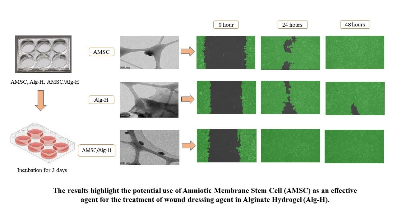
Fitriani N, Wilar G, Narsa AC, Elamin KM, Wathoni N
Stem Cells and Cloning: Advances and Applications 2025, 18:1-13
Published Date: 10 January 2025
Gallic Acid Nanocrystal Hydrogel: A Novel Strategy for Promoting Wound Healing and Inhibiting Scar Formation

Yan M, Huang S, Li X, Wang Y, Zhong S, Ban J, Zhang S
International Journal of Nanomedicine 2025, 20:4607-4626
Published Date: 11 April 2025